Foot Bones Drawing
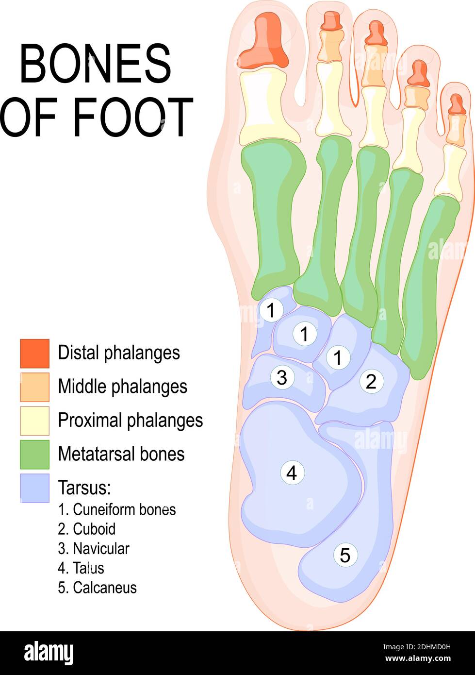
Foot Bones Drawing - The skeletal structure of the foot is. The bones in the foot make up nearly 25% of the total. Tarsals make up a strong weight bearing platform. 178k views 5 years ago anatomy of the human body for artists | proko. Bones of the foot and ankle joint medical vector illustration. These bones give structure to the foot and allow for all foot movements like flexing the toes and ankle, walking, and running. First, draw a circle for the head. Web how to draw a skeleton: In humans, the foot is one of the most complex structures in the body. Let’s look briefly at the structure of the foot: Dorsal view of the right foot, showing major muscles, tendons, and nerves. In humans, the foot is one of the most complex structures in the body. These bones give structure to the foot and allow for all foot movements like flexing the toes and ankle, walking, and running. They are homologous to the carpals in the wrist and are divided. In this video part, you will discover the complex structure of the foot bones, which a figurative fine artist must know in order to depict a foot realistically. Web the foot can also be divided up into three regions: First, draw a circle for the head. The foot is challenging to draw because it’s flexible, asymmetrical, and should usually look. More than 100 muscles, tendons, and ligaments. Side view of skeleton leg with phalange, metatarsal, tarsal and calcaneus, cuneiform, navicular and tibia bones diagram. Have a close look at the human foot anatomy. Muscles, tendons, and nerves of the human foot. Web foot bones, sketch of human anatomy. Bones of the foot and ankle joint medical vector illustration. This cornerstone is not altered as in brick work, but rather moves openly between the inward also, external condyle. Foot bones vector sketch of human anatomy, orthopedics medicine design. The bones in the foot make up nearly 25% of the total. The tarsals or ankle bones in blue, the metatarsi. The foot is challenging to draw because it’s flexible, asymmetrical, and should usually look like. The foot can be divided into three regions, the hindfoot, midfoot, and forefoot. Web there are 26 bones in the foot, divided into three groups: Web foot bones, sketch of human anatomy. Bones of the foot and ankle joint medical vector illustration. Bones of the foot and ankle joint medical vector illustration. Many of the muscles that affect larger foot movements are located in the lower leg. Web it’s time to learn how to draw a foot! 178k views 5 years ago anatomy of the human body for artists | proko. Web how to draw a skeleton: Web the foot can also be divided up into three regions: Bones of the foot and ankle joint medical vector illustration. Muscles, tendons, and nerves of the human foot. In this video part, you will discover the complex structure of the foot bones, which a figurative fine artist must know in order to depict a foot realistically. 178k views 5. Have a close look at the human foot anatomy. 7.8k views 4 years ago anatomy for artists. Web foot bones, sketch of human anatomy. The foot is challenging to draw because it’s flexible, asymmetrical, and should usually look like. Sketch the skeleton head and torso. First, draw a circle for the head. In humans, the foot is one of the most complex structures in the body. Web we have more than 475,000,000 assets on shutterstock.com as of november 30, 2023. Characters & creatures 3d models. Side view of skeleton leg with phalange, metatarsal, tarsal and calcaneus, cuneiform, navicular and tibia bones diagram. Let’s look briefly at the structure of the foot: More than 100 muscles, tendons, and ligaments. The tarsals or ankle bones in blue, the metatarsi or instep bones in purple, and the phalanges or toes in pink. 178k views 5 years ago anatomy of the human body for artists | proko. Add a face cross to mark where our facial. We will start this how to draw a skeleton tutorial with the usual simple base sketch. Foot, in anatomy, terminal part of the leg of a land vertebrate, on which the creature stands. Web there are 26 bones in the foot, divided into three groups: These bones give structure to the foot and allow for all foot movements like flexing the toes and ankle, walking, and running. Side view of skeleton leg with phalange, metatarsal, tarsal and calcaneus, cuneiform, navicular and tibia bones diagram. The tarsals or ankle bones in blue, the metatarsi or instep bones in purple, and the phalanges or toes in pink. Sketch the skeleton head and torso. Very little of the foot can move, so we can simplify it as shown on the right: Human foot bones front and side view anatomy. Web how to draw a skeleton: Bones of the foot and ankle joint medical vector illustration. Web basics of the foot. Web the bones of the foot are wedged together and bound by ligaments. The foot can be divided into three regions, the hindfoot, midfoot, and forefoot. Base sketch step by step. Add a face cross to mark where our facial features will go.
Foot bones anatomy Royalty Free Vector Image VectorStock

Foot Description, Drawings, Bones, & Facts Britannica
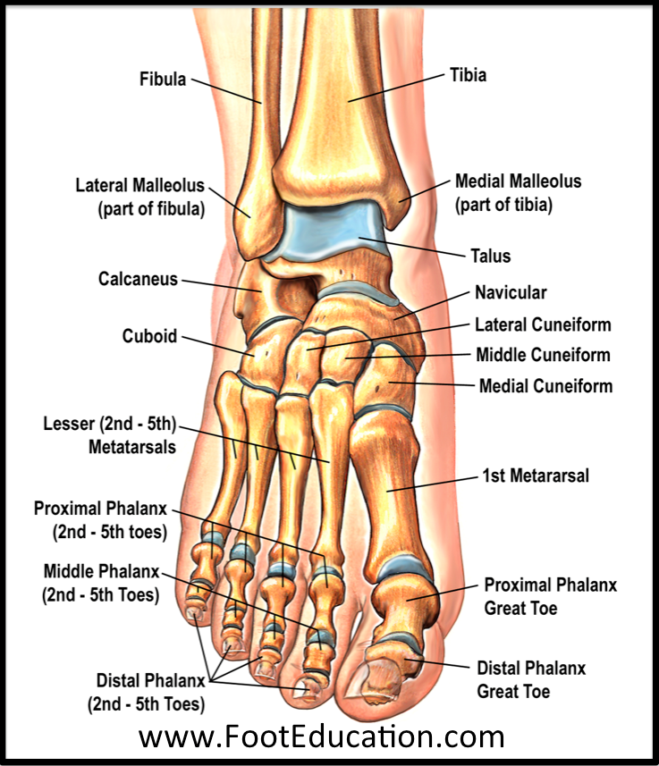
Bones and Joints of the Foot and Ankle Overview FootEducation
.jpg)
Foot Bone Diagram resource Imageshare

Bones of the Foot 2 by tiffanydavis on DeviantArt
Bones of foot. Human Anatomy. The diagram shows the placement and names
.jpg)
Foot Bone Diagram resource Imageshare

Structure of the human foot bone Royalty Free Vector Image

Foot & Ankle Bones

Foot Bone Anatomy Vector Illustration 539973 Vector Art at Vecteezy
More Than 100 Muscles, Tendons, And Ligaments.
Web It’s Time To Learn How To Draw A Foot!
Web The Foot Can Also Be Divided Up Into Three Regions:
They Are Homologous To The Carpals In The Wrist And Are Divided Into Three Groups:
Related Post: