Drawing Of Simple Squamous Epithelium
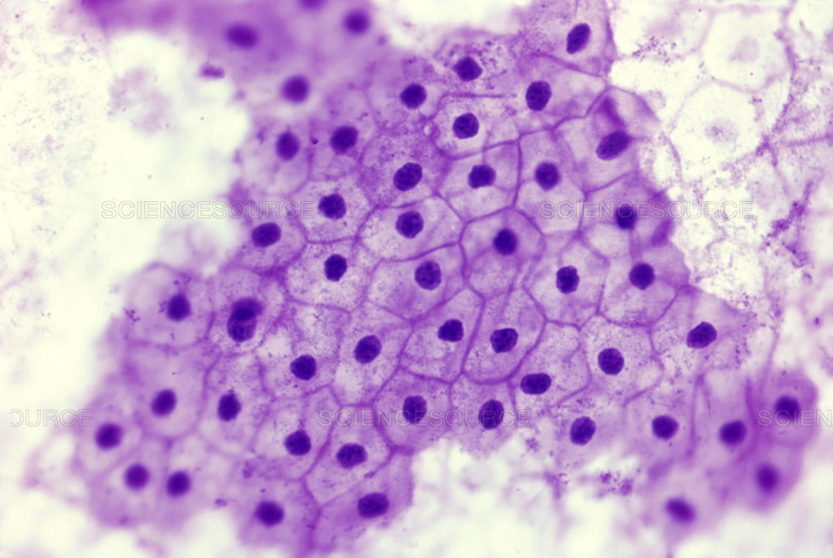
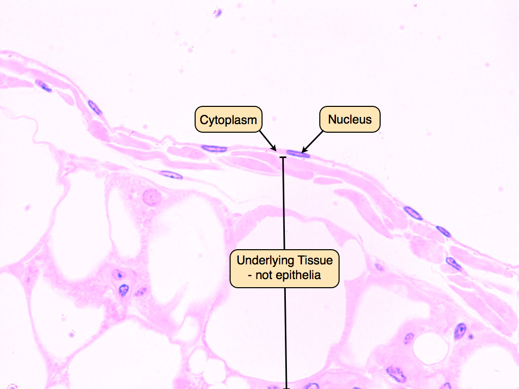
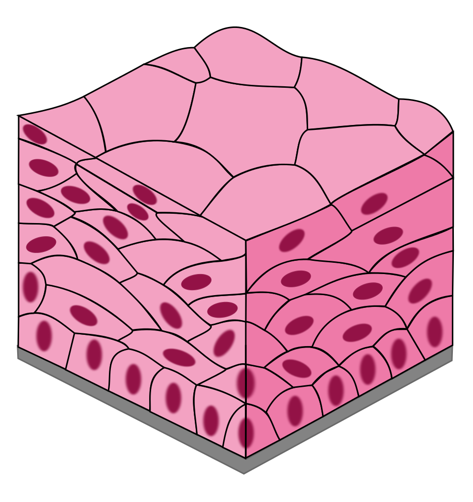
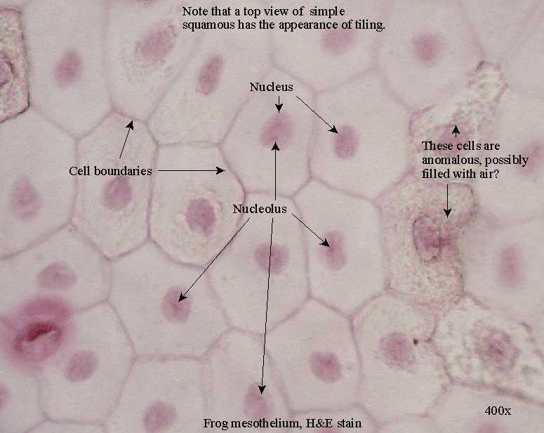
Drawing Of Simple Squamous Epithelium - However, the focus here is only on a simple squamous epithelium. Web simple squamous epithelium: Download the complete guide to neet ug prep. This is made up of thin, flat and hexagonal cells. Web simple squamous epithelium | epithelium. Web in the center of this image are two simple squamous epithelial cells that are still attached to each other. Notice that the location of the nucleus (nuc) is in the center of the cell. What are squamous epithelial cells? Renal corpuscles in the kidney consists of two structures: It is surrounded by the much paler cytoplasm (cyt). Renal corpuscles in the kidney consists of two structures: It also lines the glomeruli in the kidney and the pulmonary alveoli where passive diffusion occurs. Find one of the round structures (~250 µm diameter) known as renal corpuscles. This epithelium also lines surfaces that require minimal protection, as shown here. Web a simple squamous epithelium, also known as pavement epithelium. Web what is simple squamous epithelium? It is a type of epithelium formed by a single layer of squamous or flat cells present on a thin extracellular layer, called the basement membrane. Web a simple squamous epithelium, also known as pavement epithelium and tessellated epithelium, is a single layer of flattened, polygonal cells in contact with the basal lamina (one. A large central rounded nucleus contain by each cell. Web simple squamous epithelium | epithelium. Web what is simple squamous epithelium? 2.6k views 3 years ago pakistan. Learn about its location in the body, cells, and characteristics. What are squamous epithelial cells? Web simple squamous epithelium is composed of a single layer of thin cells that are much wider than they are tall. The cells found in this epithelium type are flat and thin, making simple squamous epithelium ideal for lining areas where passive diffusion of gases occur. 2.6k views 3 years ago pakistan. Each contains a. Simple epithelium can be divided into 4 major classes, depending on the shapes of constituent cells. Web simple squamous epithelium | epithelium. Learn about its location in the body, cells, and characteristics. Web simple squamous epithelium is a type of simple epithelium made up of squamous epithelial cells that lines the outer layer of the skin, endothelium, and secretory parts. To help you understand how to identify simple squamous epithelium, we have included two examples of this tissue. 2.6k views 3 years ago pakistan. Web simple squamous epithelium is composed of a single layer of thin cells that are much wider than they are tall. Learning about the major cells and tissues of the body is a central part of. Web simple squamous epithelia are tissues formed from one layer of squamous cells that line surfaces. A large central rounded nucleus contain by each cell. This epithelium also lines surfaces that require minimal protection, as shown here. Squamous cells are large, thin, and flat and contain a rounded nucleus. Describe the structure and function of endocrine and exocrine glands. Web now, let’s learn how to draw the simple squamous epithelium that finds under a microscope. Web simple squamous epithelium | epithelium. Simple epithelial tissues example 1. It also lines the glomeruli in the kidney and the pulmonary alveoli where passive diffusion occurs. This epithelium presents a minimal barrier to passive diffusion and, therefore, lines surfaces across which metabolites or. Learn about its location in the body, cells, and characteristics. Web simple squamous epithelium: Squamous cells are large, thin, and flat and contain a rounded nucleus. The cells found in this epithelium type are flat and thin, making simple squamous epithelium ideal for lining areas where passive diffusion of gases occur. Simple epithelium can be divided into 4 major classes,. Silver stain (hematoxylin counterstain) 10,159 x. Web simple squamous epithelium: Its primary function is to provide a smooth and protective surface. Simple epithelium can be divided into 4 major classes, depending on the shapes of constituent cells. Web now, let’s learn how to draw the simple squamous epithelium that finds under a microscope. An overview of prokaryotic & eukaryotic cells. Renal corpuscles in the kidney consists of two structures: Like other epithelial cells, they have polarity and contain a distinct apical surface with specialized membrane proteins. It also lines the glomeruli in the kidney and the pulmonary alveoli where passive diffusion occurs. The cells found in this epithelium type are flat and thin, making simple squamous epithelium ideal for lining areas where passive diffusion of gases occur. Web in the center of this image are two simple squamous epithelial cells that are still attached to each other. What are squamous epithelial cells? Notice that the location of the nucleus (nuc) is in the center of the cell. First, you should draw the basement membrane of. Each contains a glomerulus (a tuft of capillaries) surrounded by bowman's capsule. Web simple squamous epithelia are tissues formed from one layer of squamous cells that line surfaces. Web drawing histological diagram of simple squamous epithelia.useful for all medical students.drawn by using h & e pencils Find one of the round structures (~250 µm diameter) known as renal corpuscles. Silver stain (hematoxylin counterstain) 10,159 x. A large central rounded nucleus contain by each cell. To help you understand how to identify simple squamous epithelium, we have included two examples of this tissue.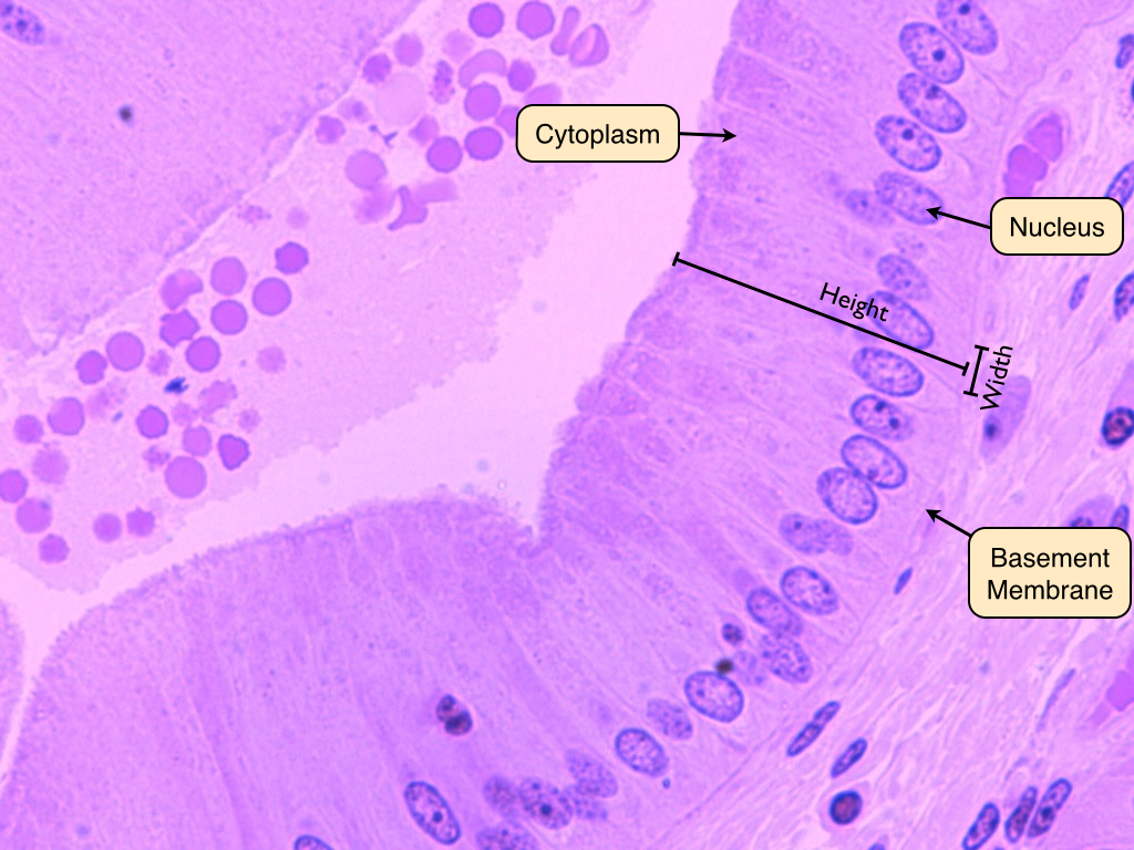
Simple Squamous Epithelium Inrtroducrion , Anatomy & Function

Transparent Body Tissues Clipart Squamous Epithelium Png Download

How to draw stratified squamous epithelium easy way YouTube
Simple Squamous Epithelium Inrtroducrion , Anatomy & Function
Epithelium Lab
Simple Columnar Epithelium Simple Squamous Epithelium Stratified
Epithelial tissues

Simple Squamous Epithelium Diagram Quizlet
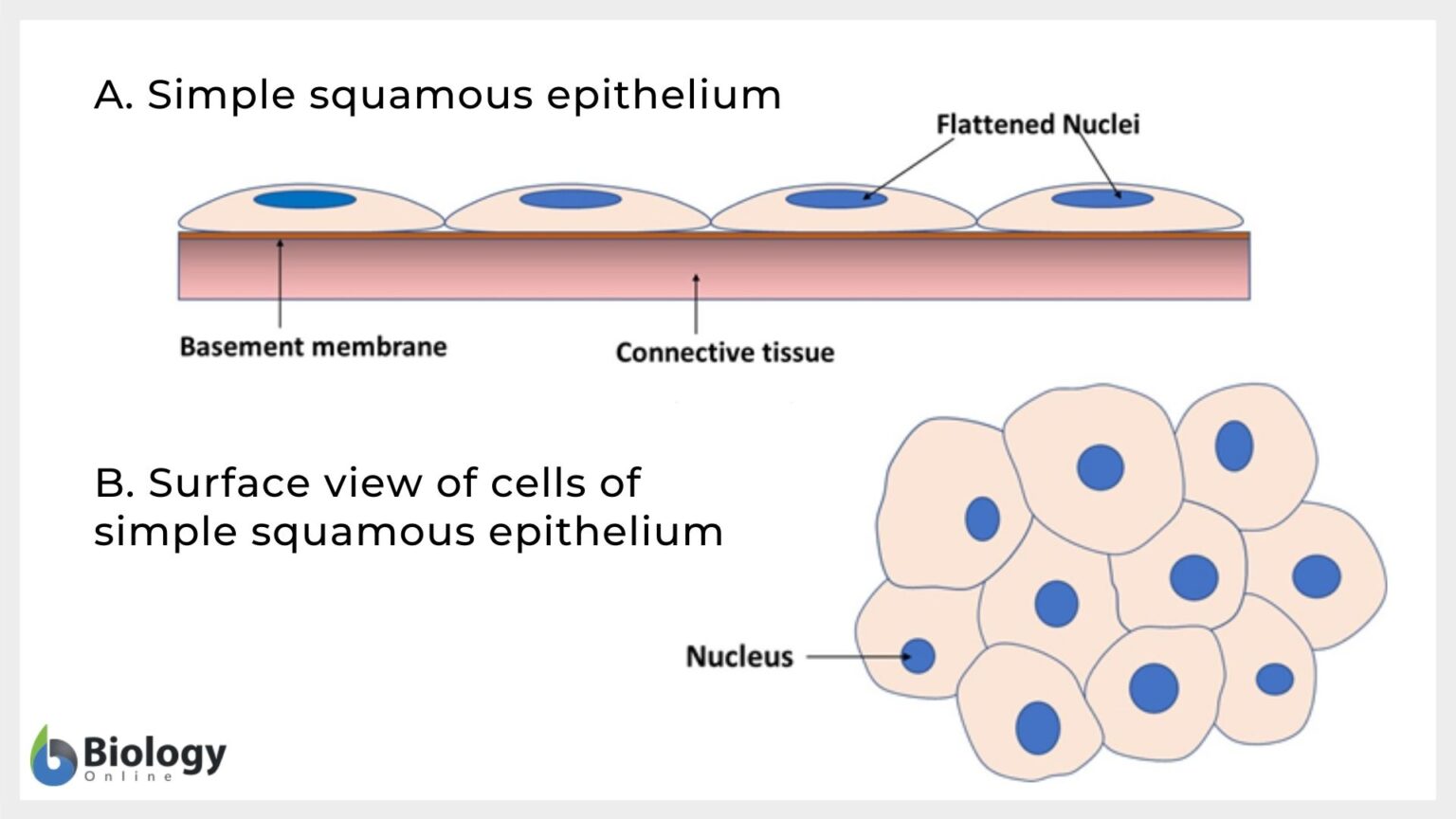
Simple squamous epithelium Definition and Examples Biology Online
Epithelial Tissue Anatomy & Physiology
Its Primary Function Is To Provide A Smooth And Protective Surface.
Web Simple Squamous Epithelium Can Be Found In Many Locations In The Body (E.g., Lining Blood Vessels, Lining The Alveoli (Air Sacs) Of Our Lungs, And In Bowman’s Capsule Of The Kidney).
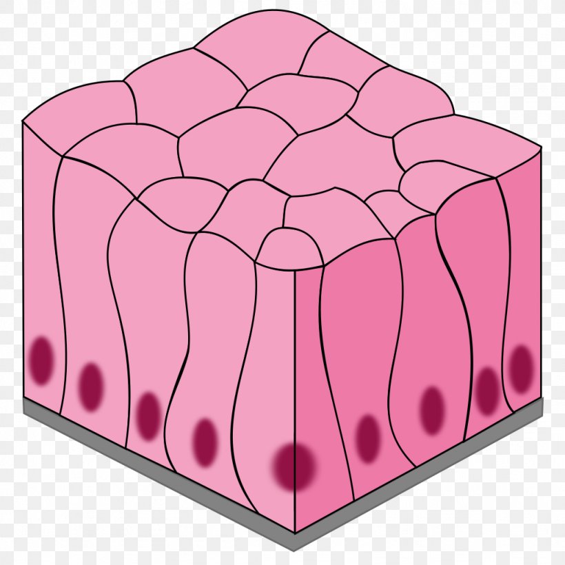
Web Now, Let’s Learn How To Draw The Simple Squamous Epithelium That Finds Under A Microscope.
Learning About The Major Cells And Tissues Of The Body Is A Central Part Of Any Biology Course.
Related Post: