Draw Sarcomere
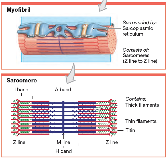
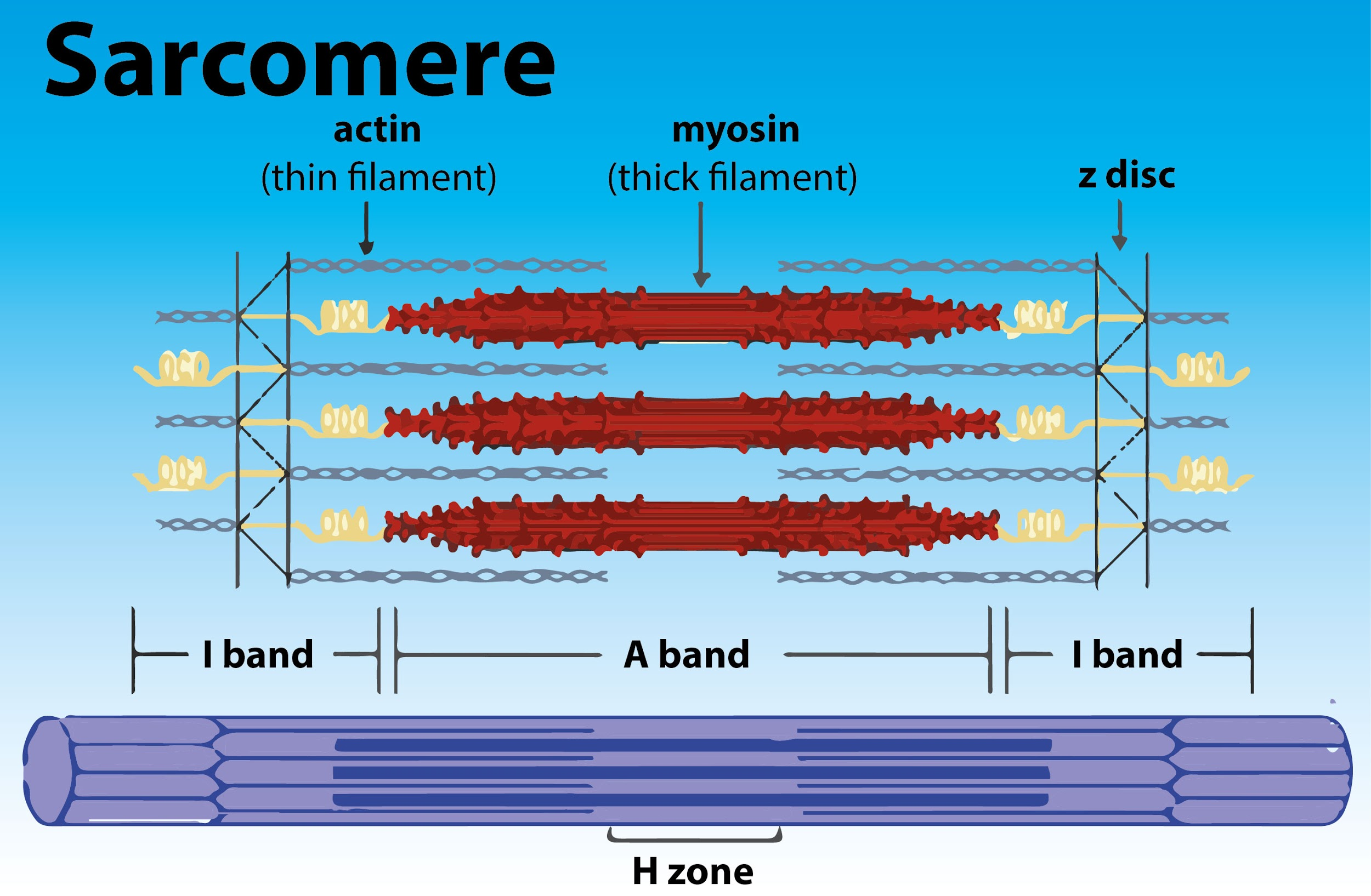
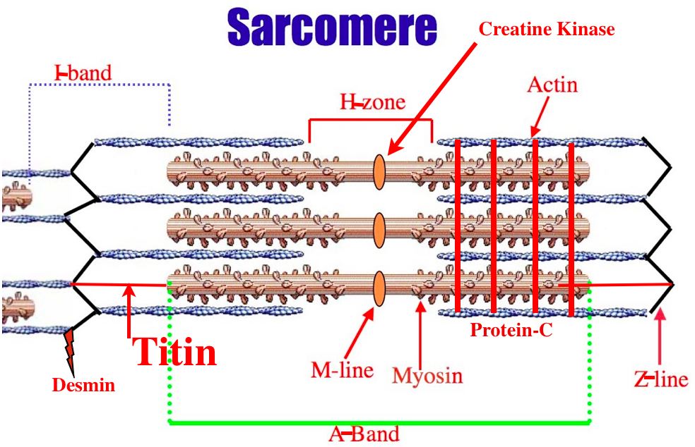
Draw Sarcomere - The sarcomere is the main contractile unit of muscle fiber in the skeletal muscle. Web as will soon be described, the functional unit of a skeletal muscle fiber is the sarcomere, a highly organized arrangement of the contractile myofilaments actin (thin filament) and myosin (thick filament), along with other support proteins. Learn vocabulary, terms, and more with flashcards, games, and other study tools. Web a sarcomere (greek σάρξ sarx flesh, μέρος meros part) is the smallest functional unit of striated muscle tissue. Muscles work on a macro level, starting with tendons that attach muscles to bones. Web about press copyright contact us creators advertise developers terms privacy policy & safety how youtube works test new features nfl sunday ticket press copyright. Hence, its main function is the regulation of muscle contraction. Web topic 11.2 skill 2 drawing labeled diagram of structure of sarcomere Each sarcomere is composed of protein filaments ( myofilaments) that include mainly the thick filaments called myosin, and thin filaments called actin. List the major sarcomeric proteins involved with contraction; Web the fundamental repeat unit within muscle that is responsible for contraction is the sarcomere. The sarcomere is the main contractile unit of muscle fiber in the skeletal muscle. Web start studying label the sarcomere structure. Explain the sliding filament process of muscle contraction Web sarcomeres are contractile units of skeletal muscle that divide into “i” and “a” bands, “m”. Hence, its main function is the regulation of muscle contraction. Some important considerations are as follows: Skeletal muscle is the muscle type that initiates all of our voluntary movement. Web as will soon be described, the functional unit of a skeletal muscle fiber is the sarcomere, a highly organized arrangement of the contractile myofilaments actin (thin filament) and myosin (thick. 8.8k views 2 years ago neet confusion series. Explain the sliding filament process of muscle contraction Some important considerations are as follows: Web by rabiya | august 10, 2019. These layers cover muscle subunits, individual muscle cells, and myofibrils respectively. The actin filaments radiate out from the z discs and help to anchor the central myosin filaments in place. This pattern is formed by a series of basic units called. How to draw a diagram of diagram of sarcomere/showing i band, a band, h zone and z line / anatomy of. Web as will soon be described, the functional unit. Muscles work on a macro level, starting with tendons that attach muscles to bones. 1.4k views 2 years ago science diagrams | explained and labelled science diagrams. A sarcomere is the basic contractile unit of a myocyte (muscle fibre). It also allows us to understand the visible bands seen in the images of muscle tissue in micrographs. A sarcomere is. Muscles work on a macro level, starting with tendons that attach muscles to bones. The function of sarcolemma is to connect the basement membrane which wrapped all connective tissues. List the major sarcomeric proteins involved with contraction; These layers cover muscle subunits, individual muscle cells, and myofibrils respectively. Explain the sliding filament process of muscle contraction Web define a muscle fiber, myofibril, and sarcomere; Skeletal muscles are composed of tubular muscle cells (called muscle fibers or myofibers) which are formed during embryonic myogenesis. 8.8k views 2 years ago neet confusion series. Hence, its main function is the regulation of muscle contraction. A sarcomere is the basic contractile unit of a myocyte (muscle fibre). It also allows us to understand the visible bands seen in the images of muscle tissue in micrographs. Design and physiology of the heart. Web define a muscle fiber, myofibril, and sarcomere; The sarcomere is the main contractile unit of muscle fiber in the skeletal muscle. The actin filaments radiate out from the z discs and help to anchor the. The sarcomere is the basic contractile unit of skeletal muscle. It is made of thick and thin filaments. The recurring sarcomeres produce a striated (striped) pattern along the length of the skeletal muscle fibres. Identify the regions of the sarcomere and whether they change during contraction; The actin filaments radiate out from the z discs and help to anchor the. The function of sarcolemma is to connect the basement membrane which wrapped all connective tissues. Herein lies the sarcomere’s main purpose. Muscles work on a macro level, starting with tendons that attach muscles to bones. Cross bridges of sarcomere in skeletal muscle are made up of. Web sarcomere is the basic contractile unit of striated muscle containing actin and myosin. Design and physiology of the heart. Explain the sliding filament process of muscle contraction The recurring sarcomeres produce a striated (striped) pattern along the length of the skeletal muscle fibres. Web the fundamental repeat unit within muscle that is responsible for contraction is the sarcomere. Within muscles, there are layers of connective tissue called the epimysium, perimysium, and endomysium. Skeletal muscles are composed of tubular muscle cells (called muscle fibers or myofibers) which are formed during embryonic myogenesis. Web start studying label the sarcomere structure. Identify the regions of the sarcomere and whether they change during contraction; List the major sarcomeric proteins involved with contraction; Some important considerations are as follows: Web a sarcomere (greek σάρξ sarx flesh, μέρος meros part) is the smallest functional unit of striated muscle tissue. The sarcomere is the main contractile unit of muscle fiber in the skeletal muscle. Web topic 11.2 skill 2 drawing labeled diagram of structure of sarcomere Web each individual sarcomere is flanked by dense protein discs called z lines, which hold the myofilaments in place. Learn vocabulary, terms, and more with flashcards, games, and other study tools. The actin filaments radiate out from the z discs and help to anchor the central myosin filaments in place.
muscular biology scheme vector illustration VectorMine

I And A Bands M And Z Lines H Zone Specialized Cell Muscle
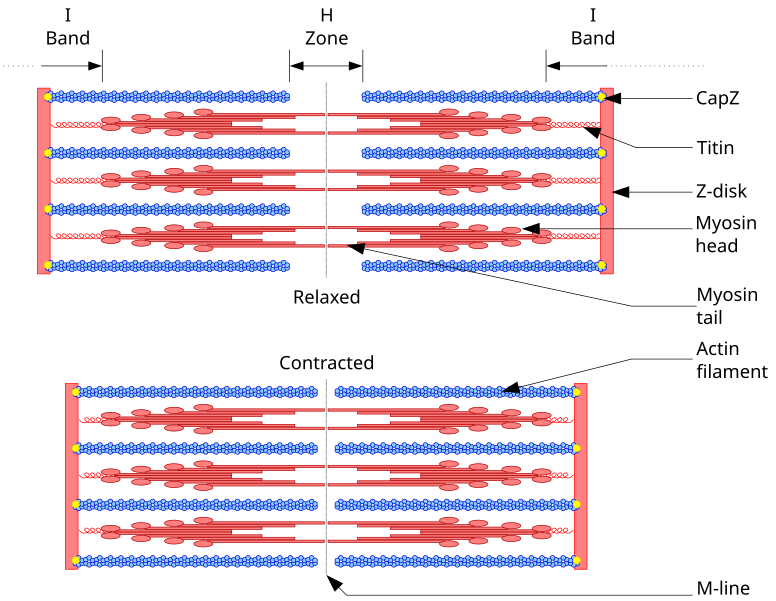
Diagram Of A

Diagram Diagram Quizlet
Definition, Structure, & Sliding Filament Theory

Contracted Diagram
Draw the diagram of a of skeletal muscle showing different
[Solved] 12. Draw and label the parts of a Course Hero

Structure of the the Contractile Unit of Skeletal Muscles
When Muscle Cells Are Viewed Under The Microscope, One Can See That They Contain A Striped Pattern (Striations).
Web Define A Muscle Fiber, Myofibril, And Sarcomere;
Web As Will Soon Be Described, The Functional Unit Of A Skeletal Muscle Fiber Is The Sarcomere, A Highly Organized Arrangement Of The Contractile Myofilaments Actin (Thin Filament) And Myosin (Thick Filament), Along With Other Support Proteins.
Each Sarcomere Is Composed Of Protein Filaments ( Myofilaments) That Include Mainly The Thick Filaments Called Myosin, And Thin Filaments Called Actin.
Related Post: