Draw Cells From The Gram Stained Slide
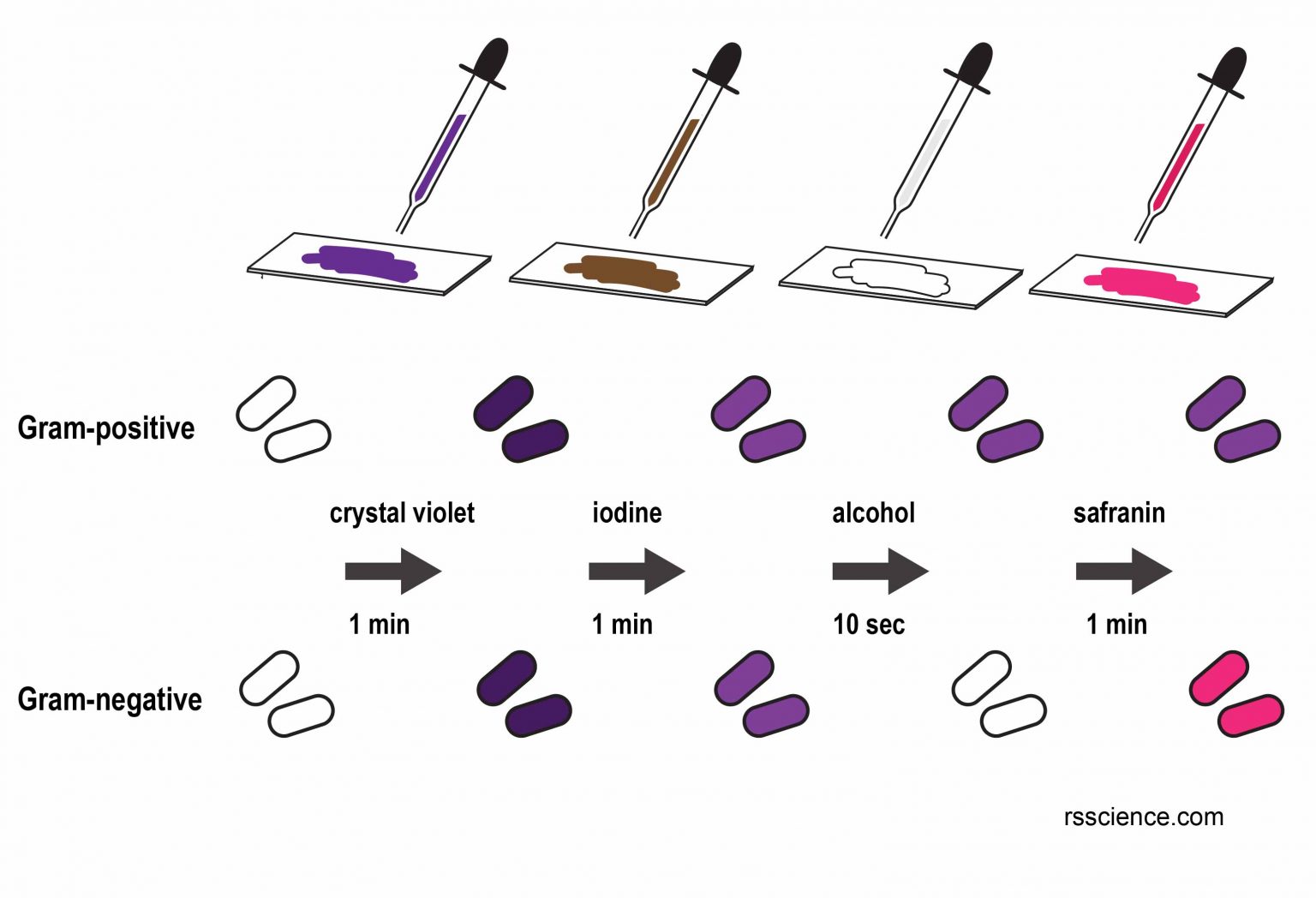
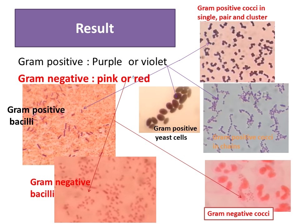
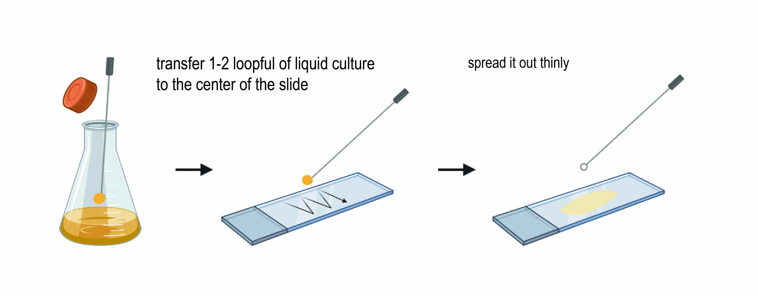
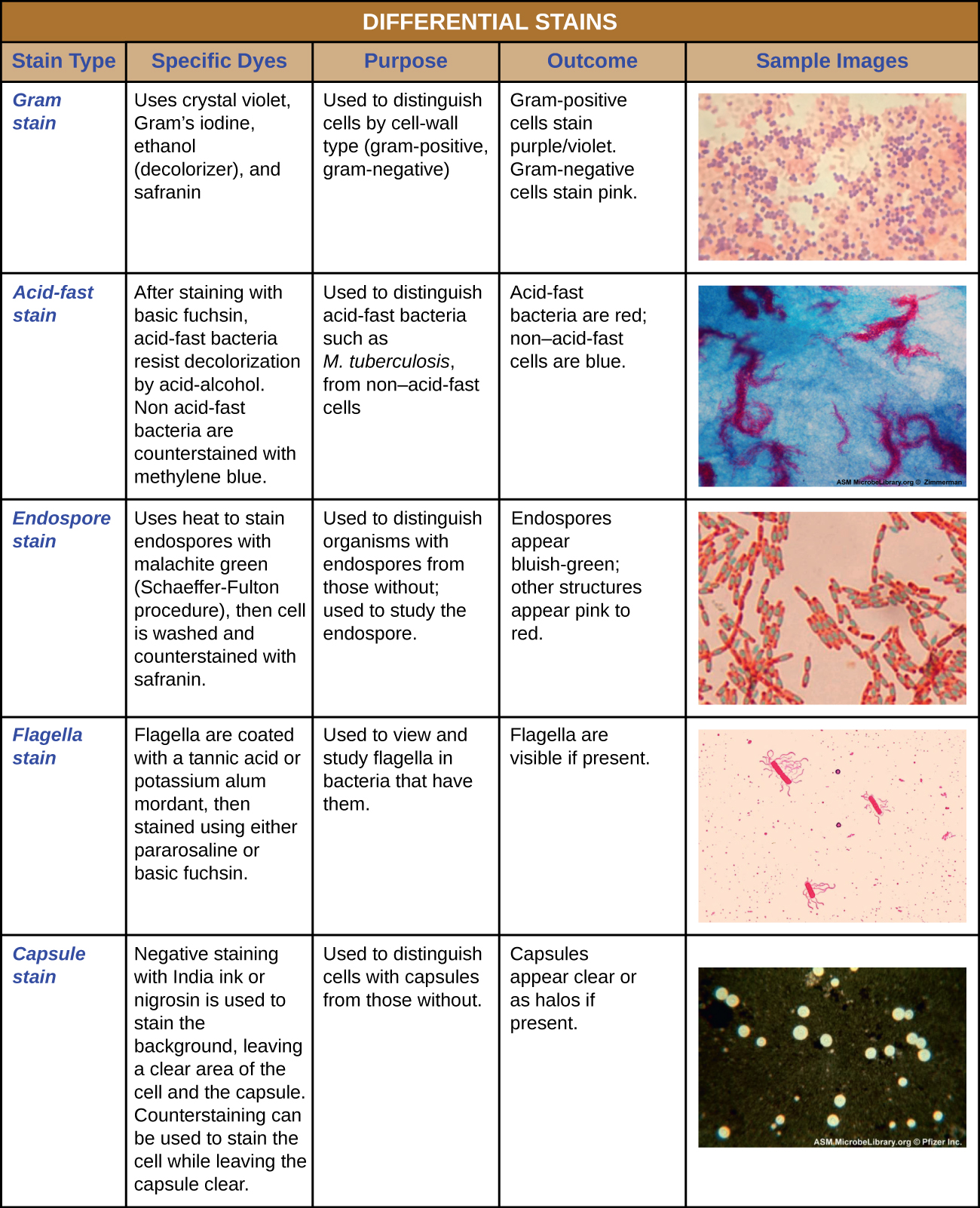
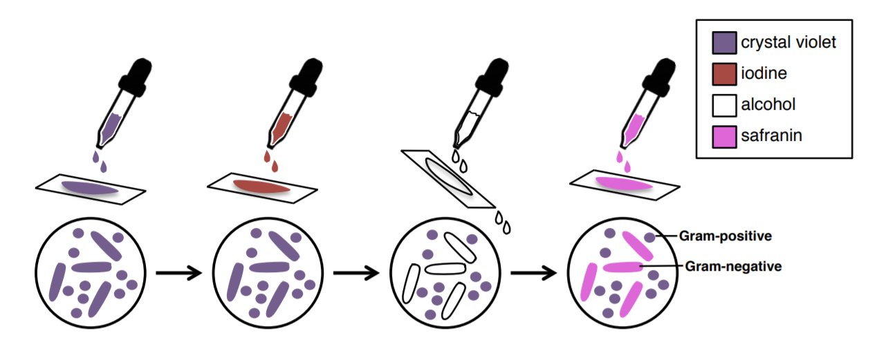
Draw Cells From The Gram Stained Slide - Web properly perform the gram staining technique. Web prepare an emulsion on each slide: Web properly perform the gram staining technique. Web gram stain procedural highlights. Web one of the most common ways to differentiate between two major groups of bacteria is using the gram stain. • quantitation of cells and microorganisms. When the bacteria is stained with primary stain crystal violet and fixed by the mordant, some of the bacteria are able to retain the primary stain and some are decolorized by alcohol. Based on your results, note the gram reaction, cell shape, and cell arrangement of each as the given table. Based on your results, note the gram reaction, cell shape, and cell arrangement of each bacterium in the given table. If you are taking a bacteria from a plate, place a small drop of water on a slide and aseptically add bacteria. Web the basic principle of gram staining involves the ability of the bacterial cell wall to retain the crystal violet dye during solvent treatment. Prepare microorganisms for microscopic observation. Explain the importance of gram stains in a clinical environment. Make sure that the liquid is completely dry before passing the slide through the flame. Explain the importance of gram stains. You and your lab partner will need to prepare the following slides: Web heating the slide briefly partially melts the cell walls, causing the cells to adhere to the glass surface. Observe the difference in size between bacteria and other unicellular microorganisms. Web rotate the 10x objective lens into place, adjust illumination if necessary, focus using the coarse adjustment, and. Staining is a key skill in microscopy work because it helps to create contrast between cells and their background. Verify these results using figure 14.5 to determine if you are correct. Web a gram stain is a laboratory test that checks for bacteria at the site of a suspected infection or in certain bodily fluids. Web 14 gram staining a.. Explain the importance of gram stains in a clinical environment. Based on your results, note the gram reaction, cell shape, and cell arrangement of each bacterium in the given table. • low power (10x) examination. Explain the function of each reagent used in a gram stain and its correlation with cell envelope structure. Based on your results, note the gram. Verify these results to p. Explain the importance of gram stains in a clinical environment. Explain the importance of gram stains in a clinical environment. Prepare microorganisms for microscopic observation. If you are taking a bacteria from a plate, place a small drop of water on a slide and aseptically add bacteria. Web properly perform the gram staining technique. Web the basic principle of the gram staining technique involves the ability of the cell wall to retain the primary stain. Student laboratory report 14 section 14 gram staining a. Explain the function of each reagent used in a gram stain and its correlation with cell envelope structure Web based on your results,. Based on your results, note the gram reaction, cell shape, and cell arrangement of each bacterium in the given table. Discover the fascinating world of bacteria! Explain the importance of gram stains in a clinical environment. Web the basic principle of gram staining involves the ability of the bacterial cell wall to retain the crystal violet dye during solvent treatment.. Identify cell morphology of bacteria. Web a gram stain is a laboratory test that checks for bacteria at the site of a suspected infection or in certain bodily fluids. Web based on your results, hote the cell shape, and cell arrangement of each bacterium in the given table. Perform a simple stain and a gram stain. Based on your results,. If you have previously viewed stained slides, you are already aware of the contrast difference made by staining techniques. Verify these results using figure 14.5 to determine if you are correct. Web the basic principle of the gram staining technique involves the ability of the cell wall to retain the primary stain. Web your gram stain slide is now ready. Discover the fascinating world of bacteria! Student laboratory report 14 section 14 gram staining a. Web the basic principle of gram staining involves the ability of the bacterial cell wall to retain the crystal violet dye during solvent treatment. Verify these results to p. A good area will look somewhat webby. Student laboratory report 14 section 14 gram staining a. Verify these results to determine if you are correct. You and your lab partner will need to prepare the following slides: Verify these results to determine if you are correct. Verify these results using figure 14.5 to determine if you are correct. If you are taking a bacteria from a plate, place a small drop of water on a slide and aseptically add bacteria. Make sure that the liquid is completely dry before passing the slide through the flame. A good area will look somewhat webby. Based on your results, note the gram reaction, cell shape, and cell arrangement of each bacterium in the given table. Verify these results to p. Explain the importance of gram stains in a clinical environment. Based on your results, note the gram reaction, cell shape, and cell arrangement of each bacterium in the given table. Typical results seen with 1000x oil immersion lens include: Web one of the most common ways to differentiate between two major groups of bacteria is using the gram stain. Prepare microorganisms for microscopic observation. Perform a simple stain and a gram stain.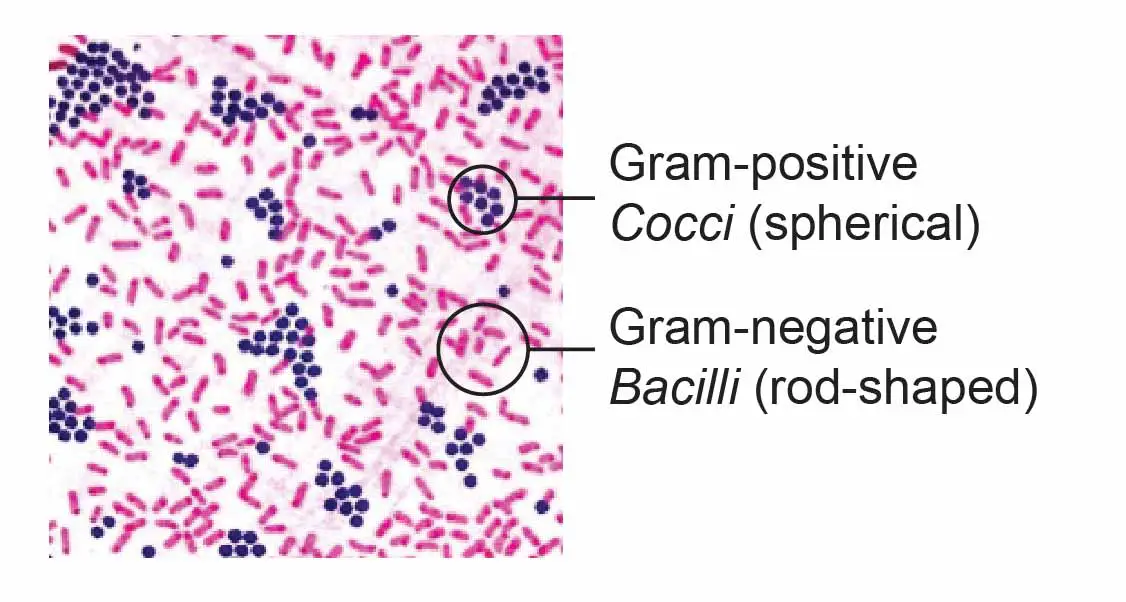
Observing Bacteria Under the Microscope Gram Stain Steps Rs' Science
Observing Bacteria Under the Microscope Gram Stain Steps Rs' Science
[Solved] GramStain technique Explain why you do each of the steps, and
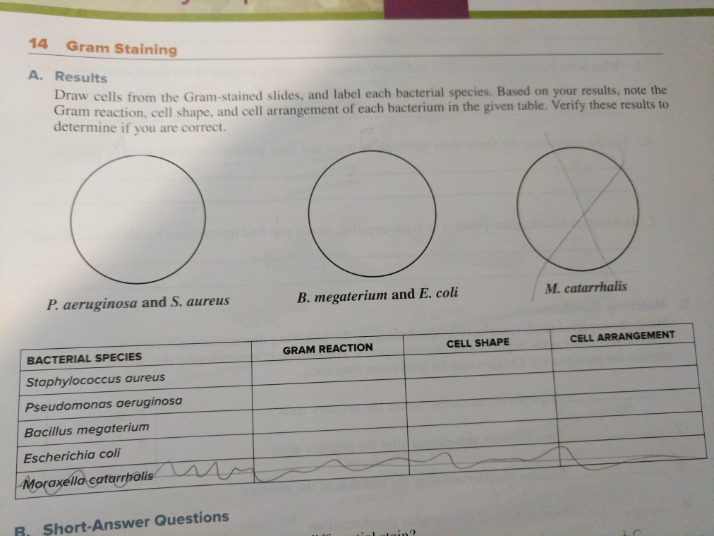
Solved 14 Gram Staining A. Results Draw cells from the

Gram Staining Principle Procedure and Results
Gram Stain Introduction, Principle, Procedure, Result and Interpretation
Observing Bacteria Under the Microscope Gram Stain Steps (2023)
1.10 Gram Stain Biology LibreTexts

Gram Staining Principle, Procedure, Results • Microbe Online
1.10 Gram Stain Biology LibreTexts
Staining Is A Key Skill In Microscopy Work Because It Helps To Create Contrast Between Cells And Their Background.
Observe The Difference In Size Between Bacteria And Other Unicellular Microorganisms.
When The Bacteria Is Stained With Primary Stain Crystal Violet And Fixed By The Mordant, Some Of The Bacteria Are Able To Retain The Primary Stain And Some Are Decolorized By Alcohol.
Web Your Gram Stain Slide Is Now Ready To Be Viewed Under The Microscope.
Related Post: