Draw A Microscope And Label
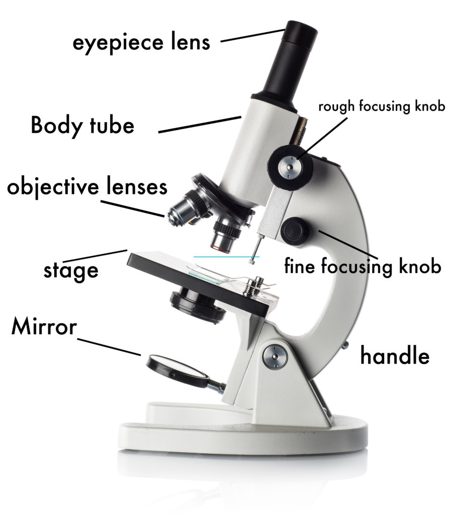
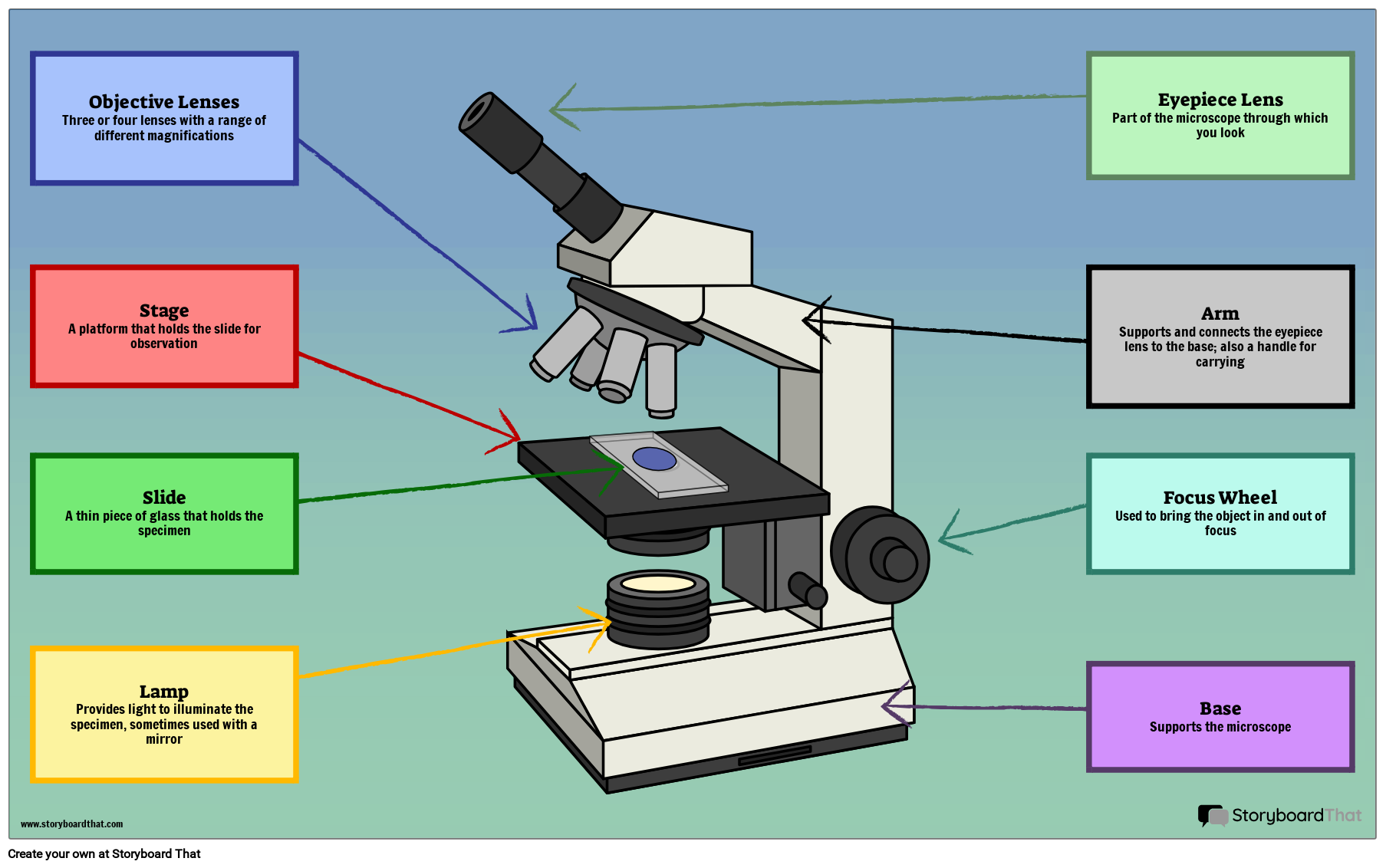
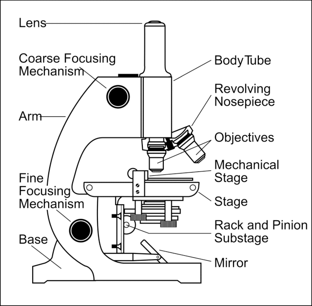
Draw A Microscope And Label - This example doesn't show the head as clearly as other microscope pictures do, so to do yours better look at a few other microscope images. Label the cell wall, cell membrane, cytoplasm, and chloroplasts in your lab manual. 6.4k views 1 year ago #microscope #howtodraw #adimushow. Be sure to indicate the magnification used and specimen name. Also indicate the estimated cell size in micrometers under your drawing. Eyepiece (ocular lens) with or without pointer: Before starting, make sure you have all the necessary materials handy. Web labeled diagram of a compound microscope. We’ll have covered the parts of both simple and compound microscopes and their functions in this article. Web this exercise is created to be used in homes and schools. Web overview of microscope and diagram. Web compound microscope definitions for labels. 195k views 3 years ago how to draw back to school! Eyepiece (ocular lens) with or without pointer: Web labeled diagram of a compound microscope. There are six printables available. Get free printable coloring page of this drawing. Structural support that holds & connects the eyepieces to the objective lenses. Black marker (optional) draw a microscope printable pdf (see bottom of lesson) the goal is to complete a drawing of a microscope by creating each part one part at a time. Web in this interactive,. Before starting, make sure you have all the necessary materials handy. Simple microscope is a magnification apparatus that uses a combination of double convex lens to. 6.4k views 1 year ago #microscope #howtodraw #adimushow. Use this with the microscope parts activity to help students identify and label the main parts of a microscope and then describe their functions. Label the. Major structural parts of a compound microscope. Label the cell wall, cell membrane, cytoplasm, and chloroplasts in your lab manual. Woman owned businessbest customer servicemicroscope rentals Web labeled diagram of a compound microscope. Use a light microscope to make observations of biological specimens and produce labelled scientific drawings. Eyepieces typically have a magnification between 5x & 30x. Using the terms listed below, label the microscope diagram. Label the cell wall, cell membrane, cytoplasm, and chloroplasts in your lab manual. Continue follow my channel and like,. Each microscope layout (both blank and the version with answers) are available as pdf downloads. #microscope #howtodraw #adimushow this is an easy and simple drawing of. By following the simple steps, you too can easily draw a perfect microscope. Today, we're learning how to draw a cool microscope! Use a light microscope to make observations of biological specimens and produce labelled scientific drawings. Woman owned businessbest customer servicemicroscope rentals It will take 9 steps in total to complete the drawing. #microscope #howtodraw #adimushow this is an easy and simple drawing of. Web this exercise is created to be used in homes and schools. 6.4k views 1 year ago #microscope #howtodraw #adimushow. Web in this interactive, you can label the different parts of a microscope. 450 views 3 years ago #chatgpt #drawing #microscope. To record the observations seen under the microscope (or from photomicrographs taken) a labelled biological drawing is often made. 6.4k views 1 year ago #microscope #howtodraw #adimushow. First and foremost, we have a labeled microscope diagram, available in both black and white and color. The microscope layout, including the blank and answered. To use a light microscope to observe, draw and label a selection of plant and animal cells, including a magnification scale. Be sure to indicate the magnification used and specimen name. It will take 9 steps in total to complete the drawing. To record the observations seen under the microscope (or from photomicrographs taken) a labelled biological drawing is often. Get free printable coloring page of this drawing. It will take 9 steps in total to complete the drawing. The part that is looked through at the top of the compound microscope. Label the cell wall, cell membrane, cytoplasm, and chloroplasts in your lab manual. Web compound microscope definitions for labels. The labeling worksheet could be used as a quiz or as part of direct instruction. Major structural parts of a compound microscope. First and foremost, we have a labeled microscope diagram, available in both black and white and color. Eyepiece (ocular lens) with or without pointer: Web overview of microscope and diagram. Attached to the tube and arm, draw the focus knob that. Label the parts of the microscope (a4) pdf print version. Label the cell wall, cell membrane, cytoplasm, and chloroplasts in your lab manual. Web this exercise is created to be used in homes and schools. 👩🎨 join our art hub. Also indicate the estimated cell size in micrometers under your drawing. #microscope #howtodraw #adimushow this is an easy and simple drawing of. Optical components of a compound microscope. Use this printable as a handout or transparency to help prepare students for working with laboratory equipment. We’ll have covered the parts of both simple and compound microscopes and their functions in this article. Black marker (optional) draw a microscope printable pdf (see bottom of lesson) the goal is to complete a drawing of a microscope by creating each part one part at a time.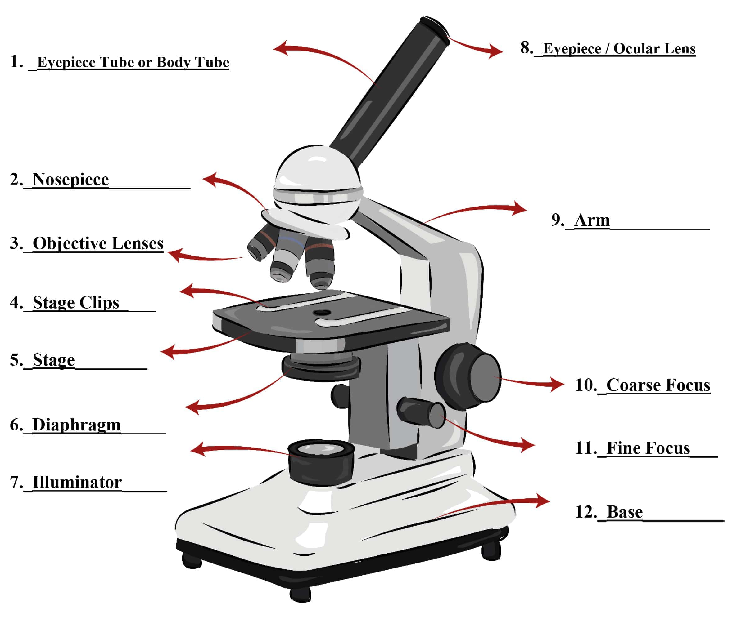
Parts of a Microscope SmartSchool Systems

Labeled Microscope Diagram Tim's Printables

Drawing Tutorial How to Draw a Microscope with Labels YouTube
Easy Microscope Labeled Diagram Micropedia vrogue.co

Simple Microscope Drawing at GetDrawings Free download
Parts of a Microscope Labeling Activity
Simple Microscope Definition, Principle, Parts, And Uses » Microscope Club

Parts of a microscope with functions and labeled diagram

Light Microscope Definition, Principle, Types, Parts, Labeled Diagram

Microscope Diagram Labeled, Unlabeled and Blank Parts of a Microscope
Today, We're Learning How To Draw A Cool Microscope!
This Example Doesn't Show The Head As Clearly As Other Microscope Pictures Do, So To Do Yours Better Look At A Few Other Microscope Images.
Web In This Interactive, You Can Label The Different Parts Of A Microscope.
Continue Follow My Channel And Like,.
Related Post: