Cheek Cell Drawing
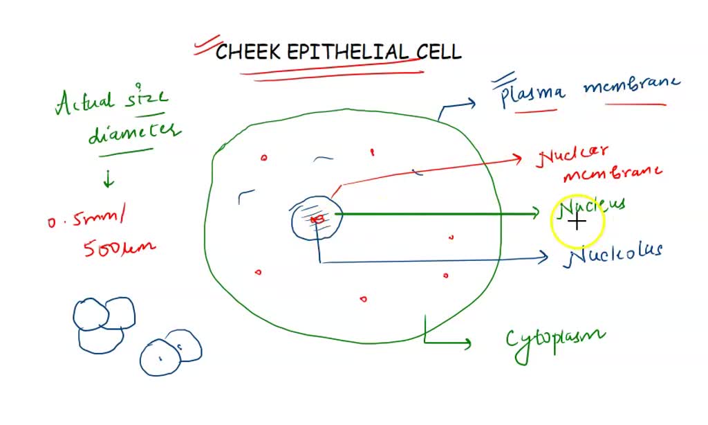
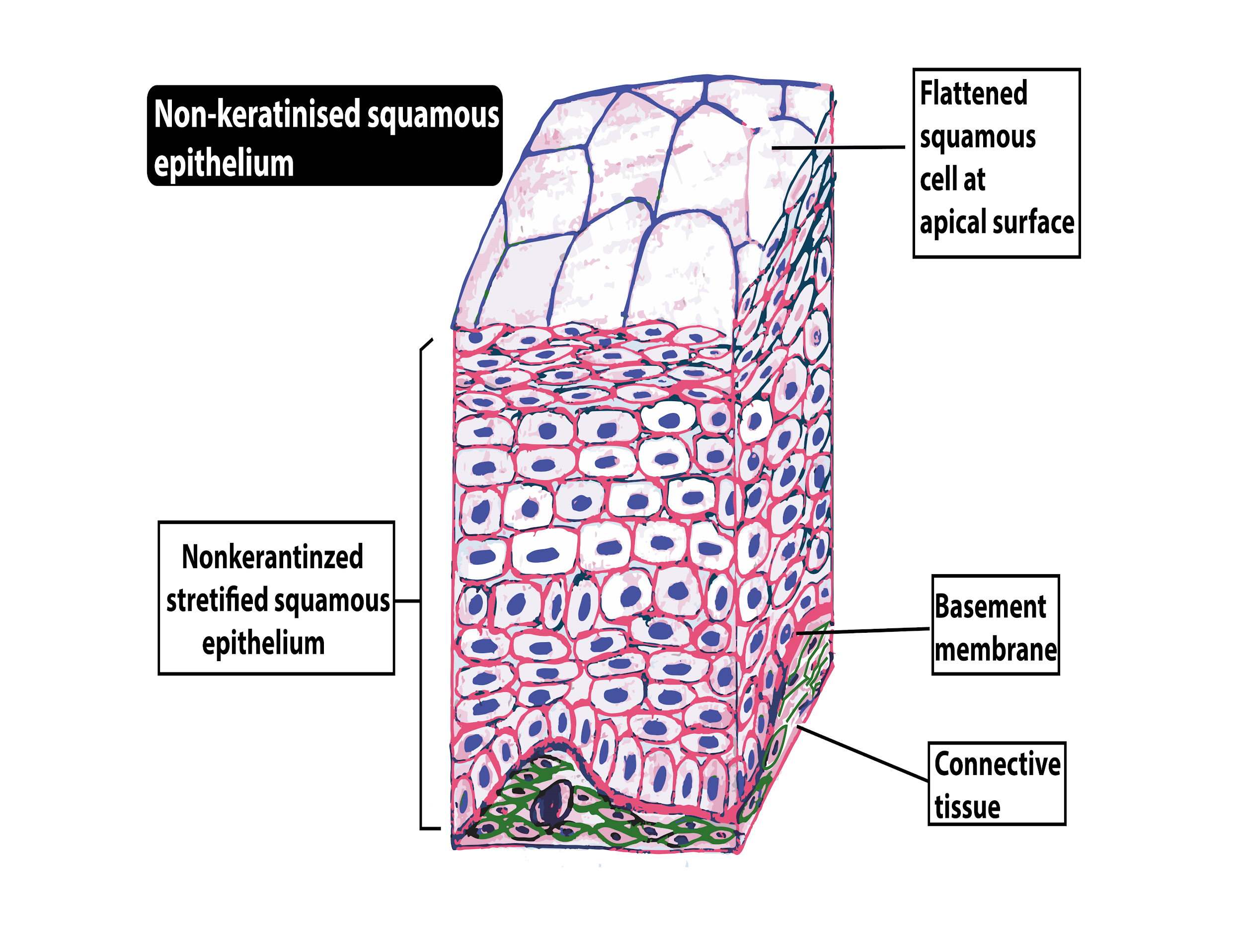
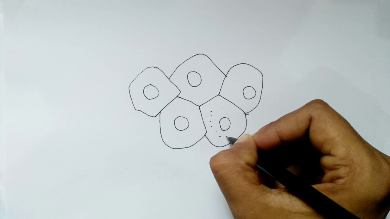
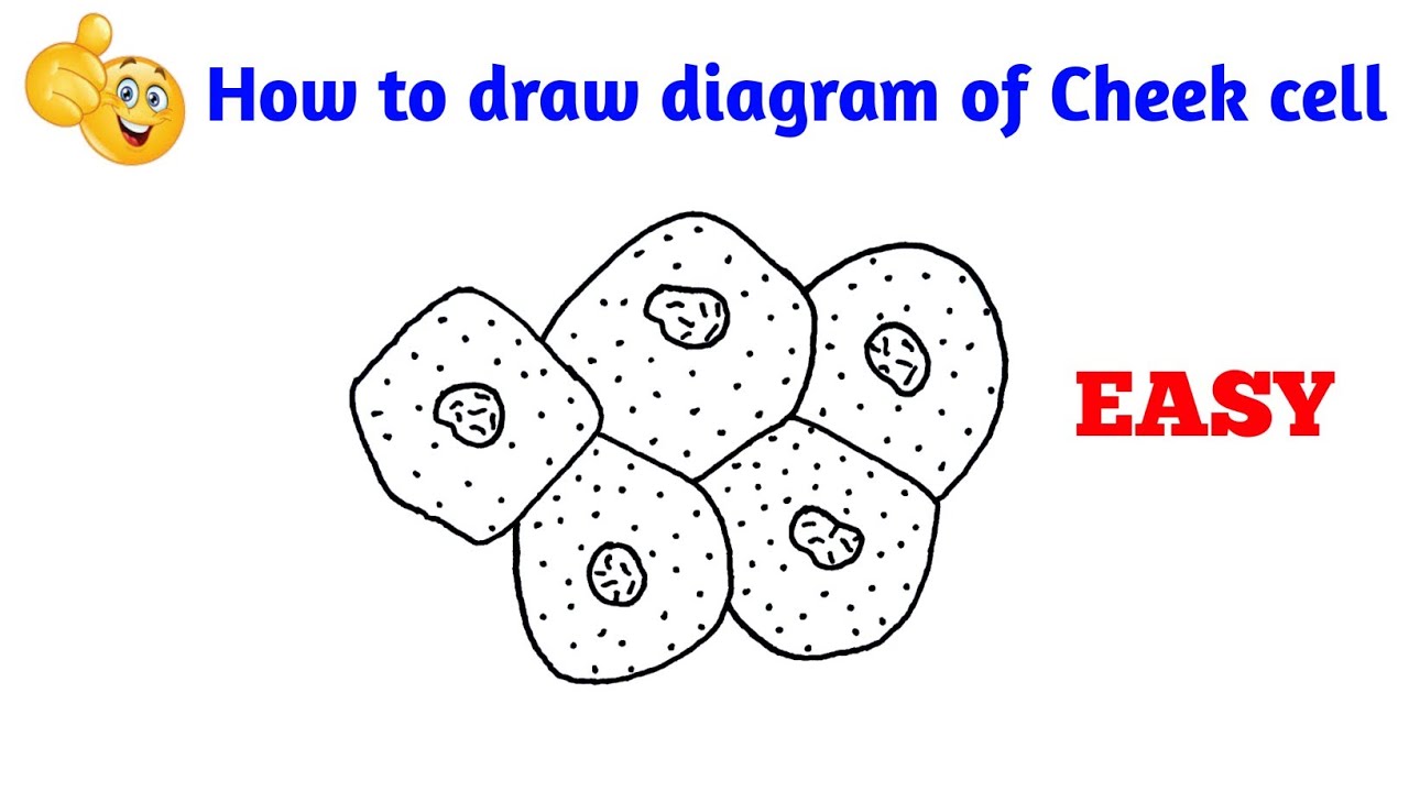
Cheek Cell Drawing - Web after watching this video you will be able to draw diagram of cheek cell in examination by using easy way for beginners. These structures, commonly thought of as cheek cells, divide approximately every 24 hours and are constantly shed from the body. Observe the cheek cells under low and high power of your microscope (at the minimum you should observe the cell membrane, nucleus, and cytoplasm). Web the human cheek cell. Draw a labelled diagram of human female reproductive system. Osmosis in potato cells takes 60 minutes after the experiment is set up. Web human cheek epithelial cells. 532 views 10 months ago #cell #diagram. Squamous epithelium is composed of thin and flat cells, with closely packed nuclei. Label the nucleus, cytoplasm, and cell membrane of a single cell. Observe the cheek cells under low and high power of your microscope (at the minimum you should observe the cell membrane, nucleus, and cytoplasm). Web draw the cheek cells at three different magnifications. Cells are stained with methylene blue and viewed with a microscope. Web (brightfield) human cheek epithelial cells at 200x magnification. How to draw human cheek cell in. List the 3 parts of the cell theory. Simple activity for observing cells. Place a cover slip on the suspension and view at 1000x total magnification. Web draw a starfish egg with a diameter of approximately 2 cm. Why is methylene blue necessary? The methylene blue was required in order to help distinguish the. The light microscope used in the lab is not powerful enough to view other organelles in the cheek cell. Study a typical animal cell to compare to your cheek cell. Web gently scrape the inside of your cheek with a toothpick and swirl it in the dye on the. The following parts of this lab should be done first because they will require time after they are set up. Label its cell membrane, cytoplasm and nucleus. The tissue that lines the inside of the mouth is known as the basal mucosa and is composed of squamous epithelial cells. Web the human cheek cell. 2.1k views 2 years ago #biology. Draw the images you see under the microscope and label the parts. 516 views 2 years ago. Web draw the cheek cells at three different magnifications. List the 3 parts of the cell theory. ∙this type of epithelium is found in the lining of the mouth and nasal cavities, blood vessels, and lymph vessels. What parts of the cell were visible. Why is methylene blue necessary? Label the nucleus, cytoplasm, and cell membrane. Compare to a plant cell investigation. Why is methylene blue necessary? Why is methylene blue necessary? , , , , , , , , , students use a toothpick to get a sample of cells from the insides of their cheek. (oblique illumination) these cells secrete mucin, a mucopolysaccharide that is the principal constituent of mucus, which helps keep the interior of the mouth moist in addition to the salivary glands.. Web after watching this video you will be able to draw diagram of cheek cell in examination by using easy way for beginners. Web can you identify the nucleus, cytoplasm and cell membrane of your cheek cell? Compare to a plant cell investigation. Click the link or scroll to go to that section of the lab. Web this lab outlines. Web this should draw the stain through and color the cells. Web human cheek epithelial cells. Web draw the cheek cells at three different magnifications. Web draw a starfish egg with a diameter of approximately 2 cm. Osmosis in potato cells takes 60 minutes after the experiment is set up. What parts of the cell were visible. Web gently scrape the inside of your cheek with a toothpick and swirl it in the dye on the slide. Web sketch the cell at low and high power. Draw your cells to scale. 532 views 10 months ago #cell #diagram. Why is methylene blue necessary? Cheek epithelial cells cells that cover a surface, whether outside the body or inside the body are called epithelial cells. 516 views 2 years ago. Why is methylene blue necessary? Sketch the cell at low and high power. These structures, commonly thought of as cheek cells, divide approximately every 24 hours and are constantly shed from the body. Web the human cheek cell. Draw the images you see under the microscope and label the parts. Web very easy way to draw human cheek cells step by step #class8#class9practicalbiology# Web (brightfield) human cheek epithelial cells at 200x magnification. The following parts of this lab should be done first because they will require time after they are set up. Web can you identify the nucleus, cytoplasm and cell membrane of your cheek cell? Squamous epithelium is composed of thin and flat cells, with closely packed nuclei. Observe the cheek cells under low and high power of your microscope (at the minimum you should observe the cell membrane, nucleus, and cytoplasm). The tissue that lines the inside of the mouth is known as the basal mucosa and is composed of squamous epithelial cells. Web how to draw human cheek cell in just 2 minutes.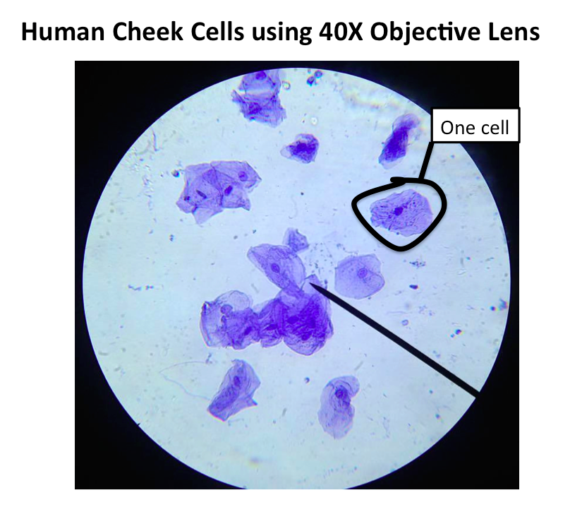
Do Human Cheek Cells Have A Nucleus Epithelial Cheek Cells Observed

Schematic Image Of A Cheek Cell

All sizes cheek cells 400x stained Flickr Photo Sharing!
SOLVED Cheek epithelial cells draw and label cell membrane, nucleus
Squamous Epithelial Cheek Cells Labeled

Do Cheek Cells Have A Nucleus / Onion Cell And Cheek Cell
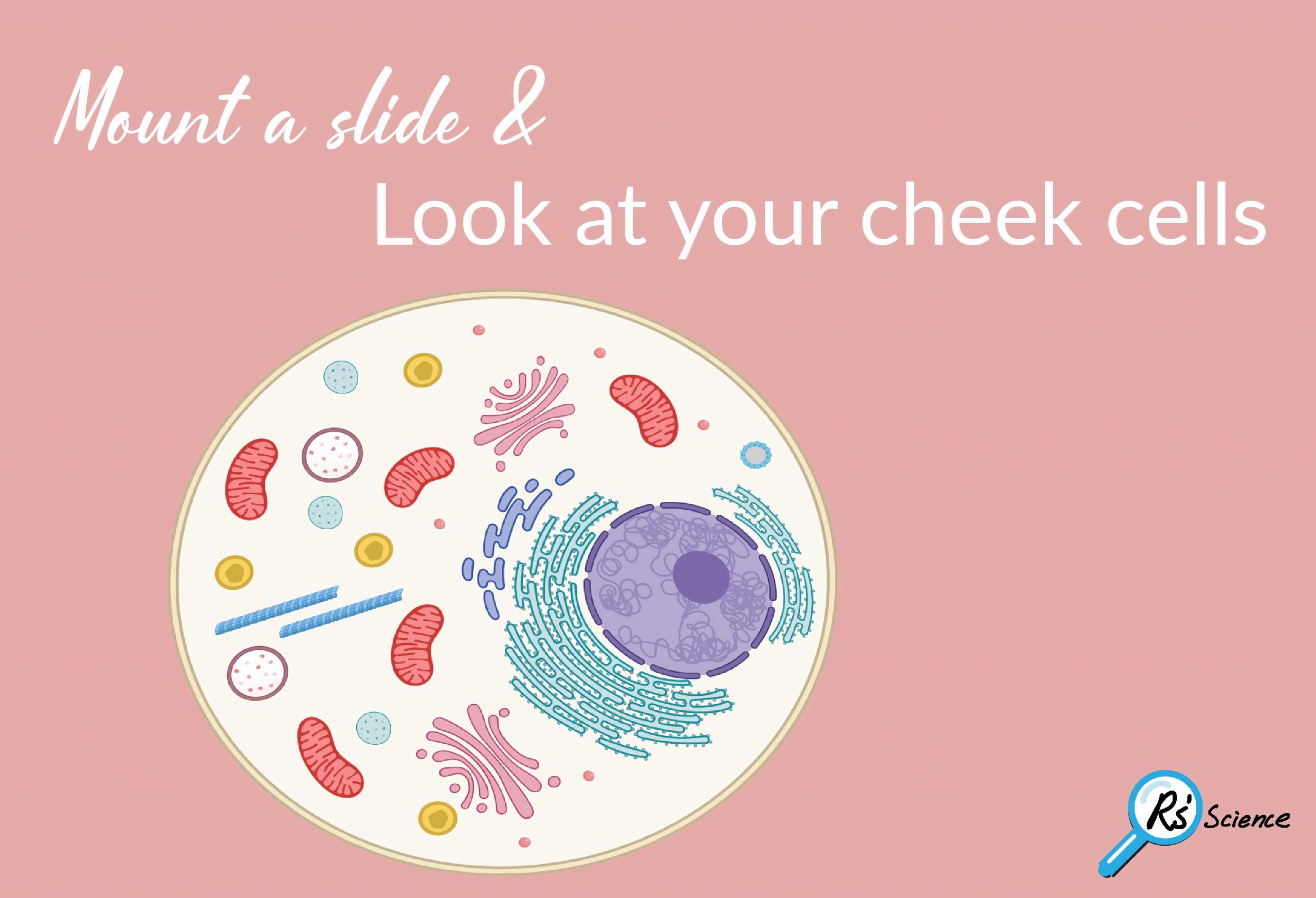
Lesson 2 Mount a Slide & “Look at Your Cheek Cells“ Rs' Science
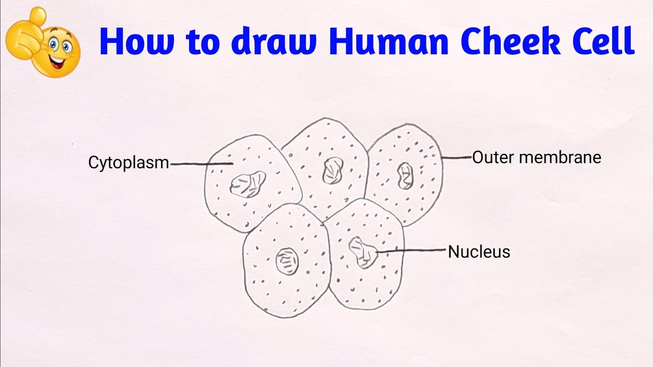
how to draw cheek cell step by step diagram of human cheek cell YouTube
How to draw Human Cheek Cell/2019 YouTube
how to draw cheek cell how to draw diagram of human cheek cell YouTube
, , , , , , , , , Students Use A Toothpick To Get A Sample Of Cells From The Insides Of Their Cheek.
The Methylene Blue Was Required In Order To Help Distinguish The.
Compare To A Plant Cell Investigation.
Web This Should Draw The Stain Through And Color The Cells.
Related Post: