Centrioles Drawing
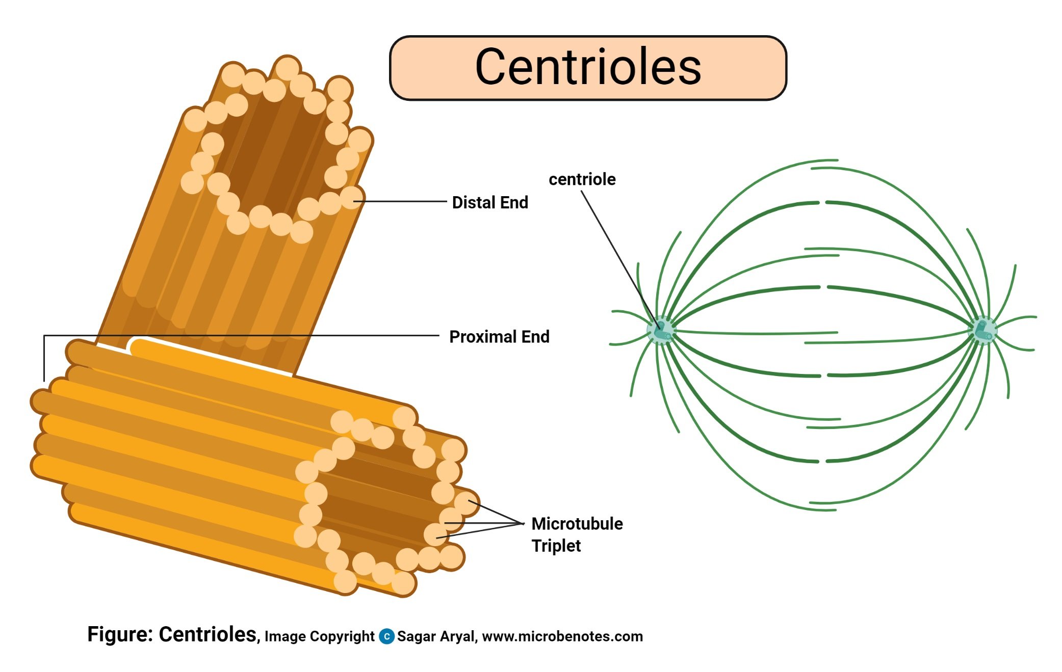
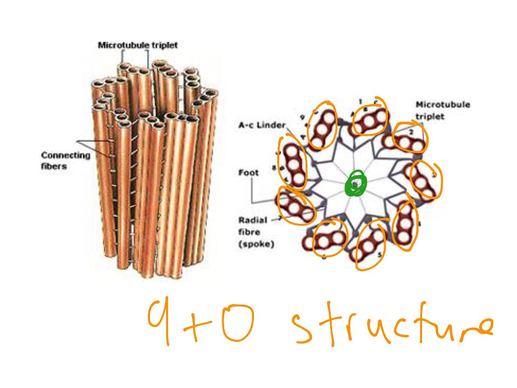
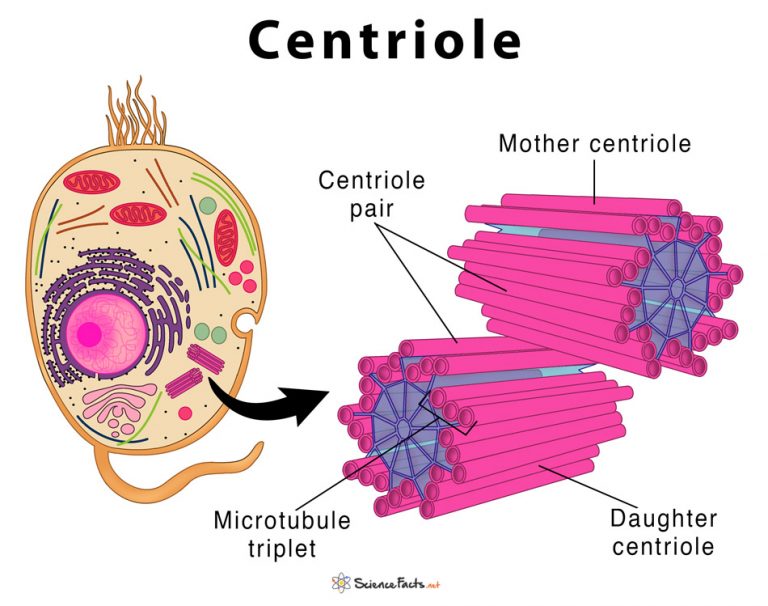
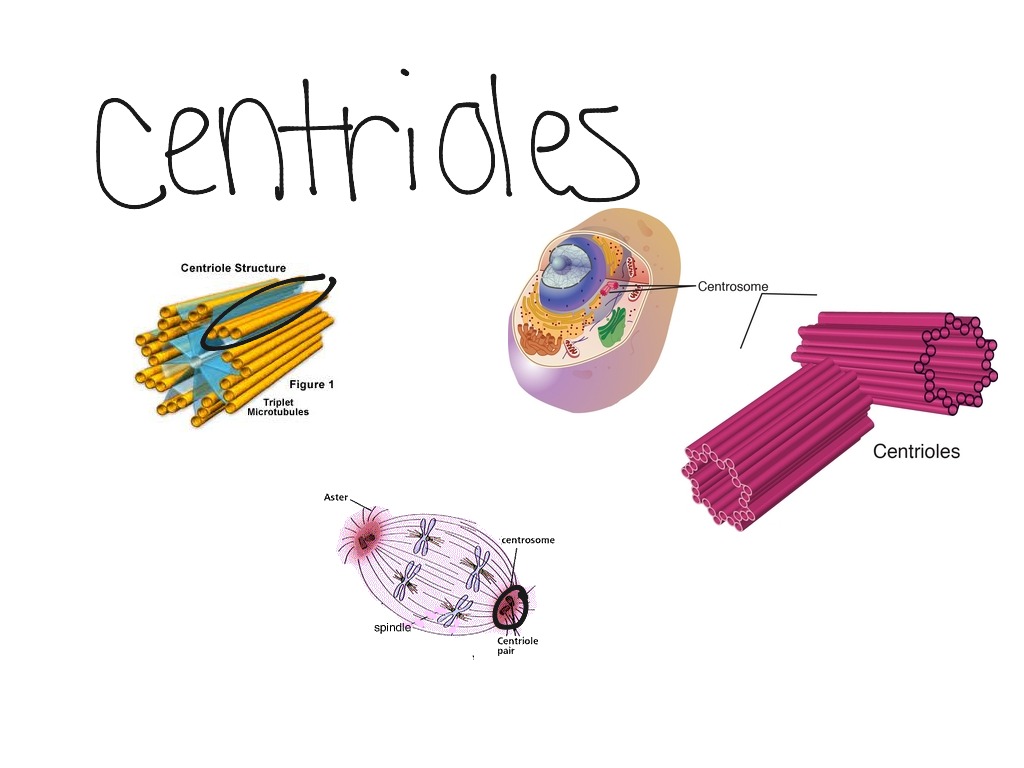
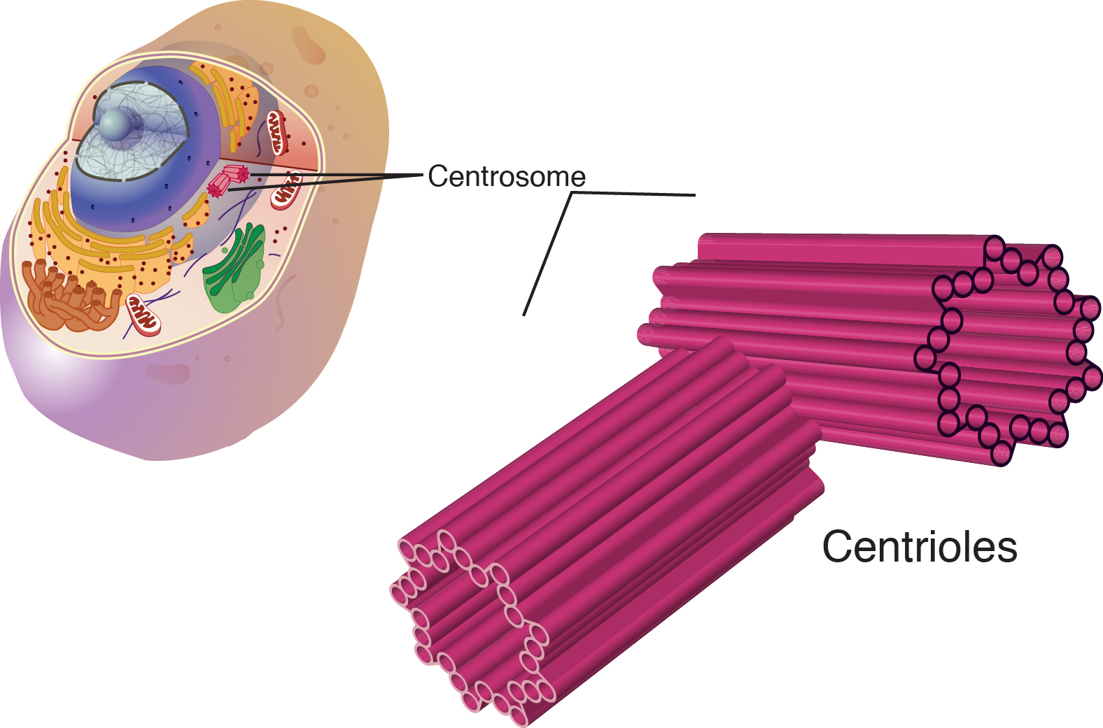
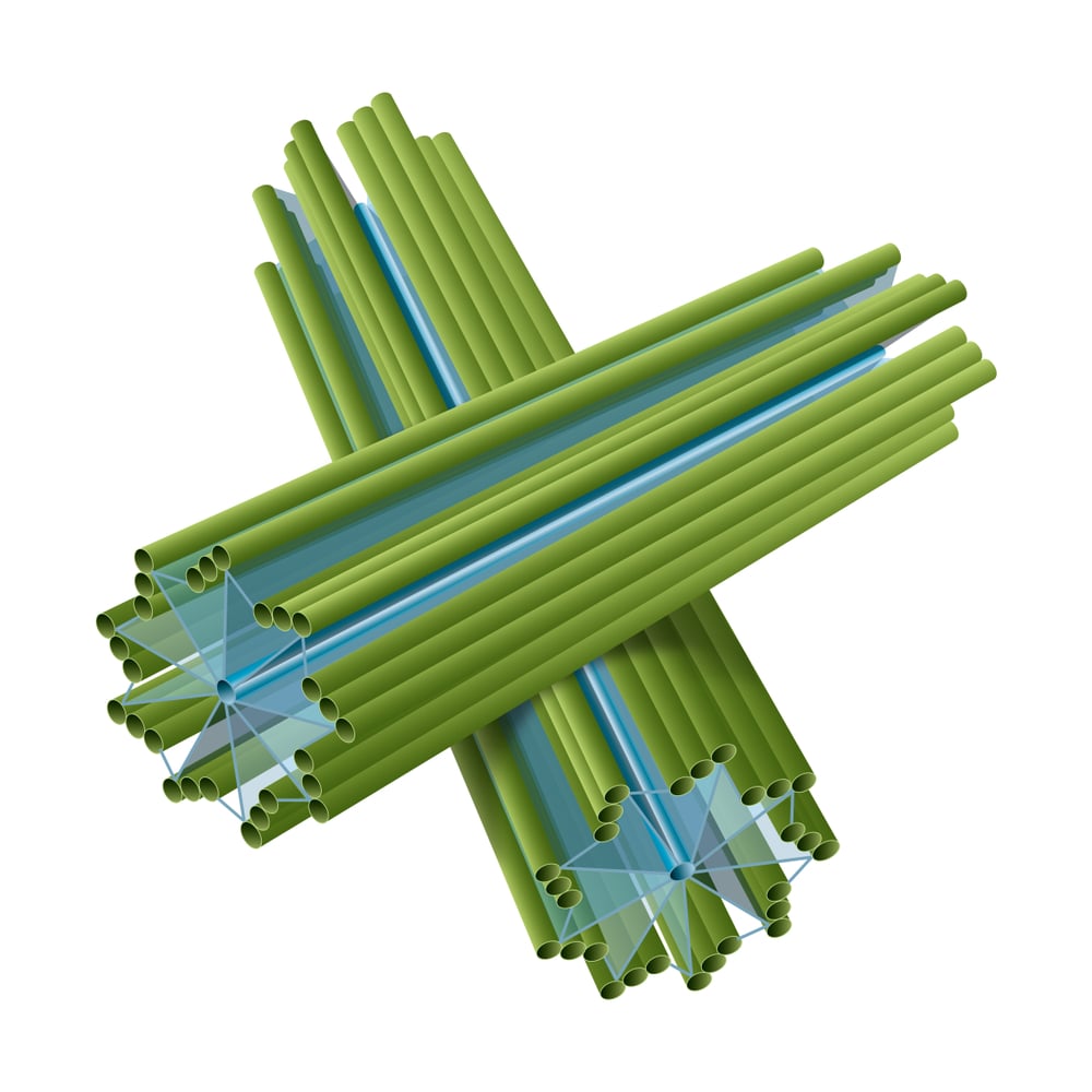
Centrioles Drawing - Deharven) is an electron micrograph showing a cross section of a centriole with its array of nine triplets of microtubules. Web centrioles are best known for their role in centrosomes, structures that act as microtubule organizing centers in animal cells. Web tufts university & harvard. It is very easy to draw. Web a centriole is a small structure made of microtubules which exists as part of the centrosome, which helps organize microtubules in the body. Within that centrosome there are two centrioles. Web the centrosome is a part of almost all plant and animal cells that includes a pair of centrioles, which are structures consisting of an array of nine microtubule triplets. Web draw two small rectangles at right angles to represent centrioles. Centrioles are built from a cylindrical array of 9 microtubules, each of which has attached to it 2 partial microtubules. You will also learn, the stepwise method for the construction of the centriole diagram. This video is regarding how to draw. 195 views 1 year ago #biology #centriole. Web draw two small rectangles at right angles to represent centrioles. The photo (courtesy of e. Web after watching this video you will be able to draw a transverse section of centriole easily in examination by using easy way for beginners. A centrosome consists of two centrioles oriented at right angles to each other, surrounded by a mass of pericentriolar material, which provides anchoring sites for microtubules 8 . Web in this animation, you will learn how to draw centriole diagram. Web tufts university & harvard. They are close to but separate from the nucleus. The photo (courtesy of e. Web in this animation, you will learn how to draw centriole diagram. Web after watching this video you will be able to draw centrioles easily in examination by using easy way for beginners. It's next to the nucleus and within the centrosome. It is very easy to draw. Centrioles are built from a cylindrical array of 9 microtubules, each of. Learn about (9+0) arrangement of. If you survived biology class, you may recall looking at grainy photos of different cell structures, such as mitochondria, nuclei, or centrioles. Web rather it appears that new centrioles are either produced de novo or are synthesized using an existing centriole as some form of template. Typically, a eukaryotic cell has one centriole that is. Learn about (9+0) arrangement of. Centrioles are built from a cylindrical array of 9 microtubules, each of which has attached to it 2 partial microtubules. Web tufts university & harvard. How to draw diagram of centriole step by step for beginnershello. Cartwheel and satellites or pericentriolar bodies. Use the pipe cleaner chromosomes to model the prophase stage of mitosis. The word some refers generally to an organelle of some sort, like a lysosome or an endosome. Centrioles help assist with cell division. Web centrioles would move toward opposite poles of the nucleus. Web after watching this video you will be able to draw centrioles easily in examination. The photo (courtesy of e. Form the centrioles by drawing two small rectangles. A centriole is the main unit that creates and anchors microtubules in the cell. Be sure to draw the cell membrane, nucleus, nucleolus, and centrioles on the paper. This video is regarding how to draw. The word some refers generally to an organelle of some sort, like a lysosome or an endosome. Learn about (9+0) arrangement of. A centriole is the main unit that creates and anchors microtubules in the cell. It's next to the nucleus and within the centrosome. Web in cell biology a centriole is a cylindrical organelle composed mainly of a protein. In the latter case, growth of the new centriole occurs at right angles to the long axis of the existing centriole, the two organelles separated from each other by a distance of 50 to 100 nm. They are close to but separate from the nucleus. This video is regarding how to draw. Centrioles are visible under a light microscope but. A centriole is the main unit that creates and anchors microtubules in the cell. You will also learn, the stepwise method for the construction of the centriole diagram. Centrioles help assist with cell division. Typically, a eukaryotic cell has one centriole that is at a right angle to a second centriole in the centrosome. Centrioles are built from a cylindrical. Web centrioles are best known for their role in centrosomes, structures that act as microtubule organizing centers in animal cells. The photo (courtesy of e. Centrioles are built from a cylindrical array of 9 microtubules, each of which has attached to it 2 partial microtubules. This video is regarding how to draw. Typically, a eukaryotic cell has one centriole that is at a right angle to a second centriole in the centrosome. Within that centrosome there are two centrioles. A centriole is the main unit that creates and anchors microtubules in the cell. Use the pipe cleaner chromosomes to model the prophase stage of mitosis. Web the centrosome is a part of almost all plant and animal cells that includes a pair of centrioles, which are structures consisting of an array of nine microtubule triplets. Cartwheel and satellites or pericentriolar bodies. Web tufts university & harvard. Web updated july 12, 2023. Be sure to draw the cell membrane, nucleus, nucleolus, and centrioles on the paper. It's next to the nucleus and within the centrosome. Deharven) is an electron micrograph showing a cross section of a centriole with its array of nine triplets of microtubules. Web in cell biology a centriole is a cylindrical organelle composed mainly of a protein called tubulin.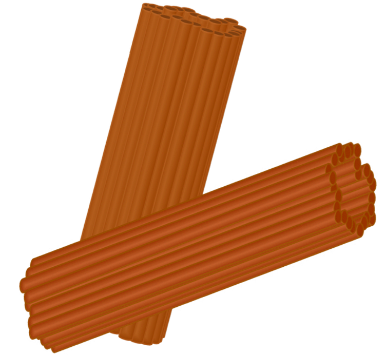
EduPic Cell Drawings
Animal Cell Definition, Structure, Parts, Functions, Labeled Diagram
Structure of centriole Science, Biology, Cell Structure ShowMe

FileCentrioleen.svg Wikimedia Commons

Centriole stock illustration. Illustration of anatomy 71079089

Centriole structure stock illustration. Illustration of mitochondria
Centriole Definition, Structure, & Functions, with Diagram
Centrioles Diagram
Cell Division II Biology Visionlearning
Centrioles Definition, Location, Role, And Function
Centrioles Are Visible Under A Light Microscope But Can Be Viewed In Detail Only Under An.
If You Survived Biology Class, You May Recall Looking At Grainy Photos Of Different Cell Structures, Such As Mitochondria, Nuclei, Or Centrioles.
How To Draw Diagram Of Centriole Step By Step For Beginnershello.
1 Is An Electron Micrograph Showing A Cross Section Of A.
Related Post: