Cardiac Muscle Drawing
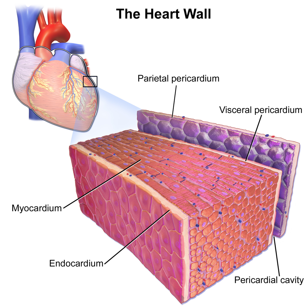
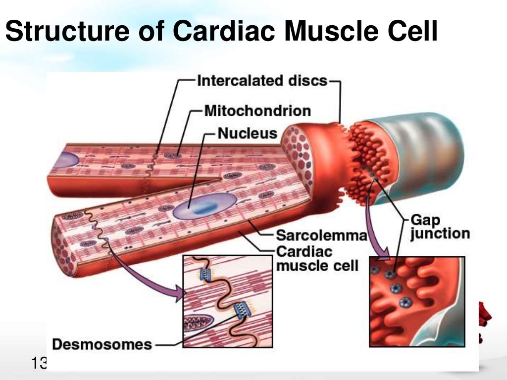
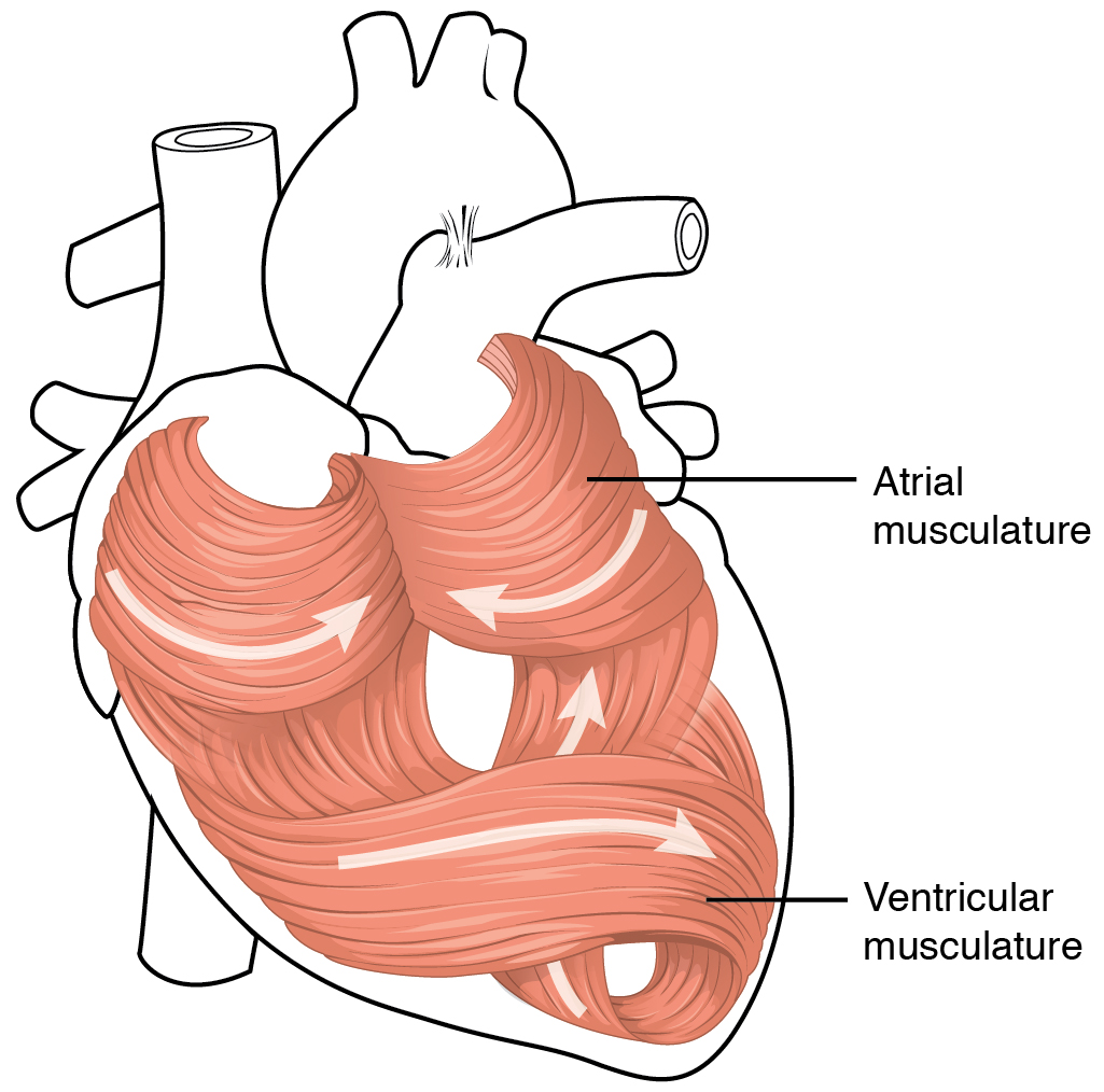
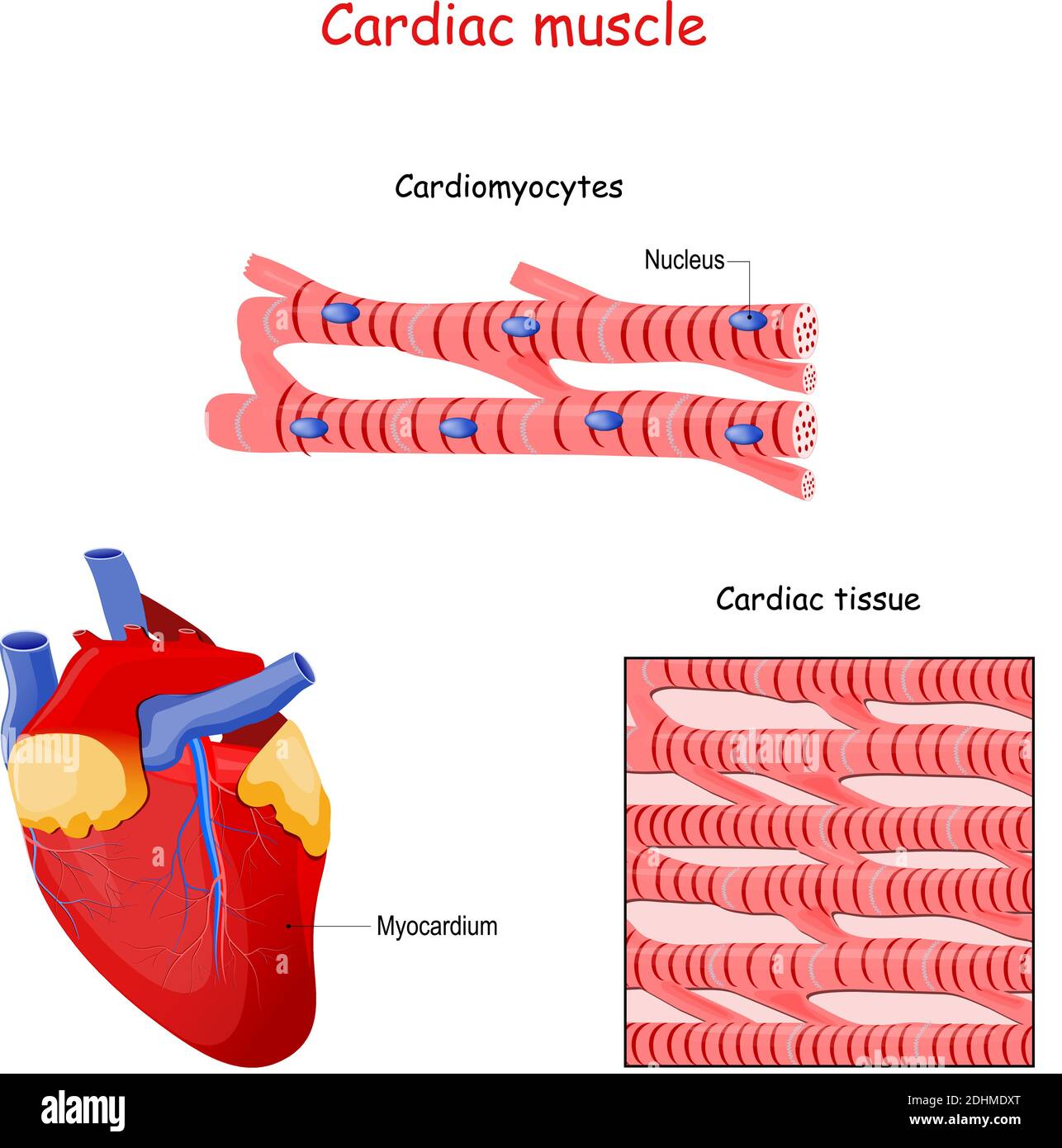
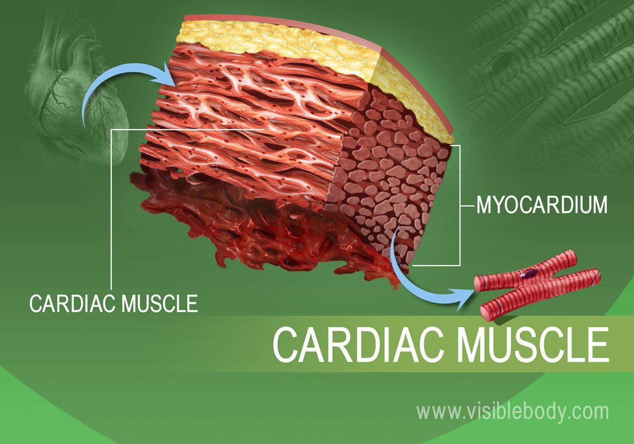
Cardiac Muscle Drawing - Heart (right lateral view) the heart is a muscular organ that pumps blood around the body by circulating it through the circulatory/vascular system. Identify and describe the components of the conducting system that distributes electrical impulses through the heart. Let's learn more about the cardiac muscle with the help of a diagram. After the end of the article, i will share the cardiac muscle histology drawing with you. Cardiac muscle tissue contracts and releases. Here, i will provide both longitudinal and transverse sectional cardiac muscle drawings. Cardiac muscle (or myocardium) makes up the thick middle layer of the heart. 1 waiting premieres may 2, 2023 #histology #anatomy #lpanatomy. Web keep exploring byju’s biology for more such exciting diagram topics. I will also enlist the functions and identification points of cardiac muscle. Similar to skeletal muscle, cardiac muscle is striated and organized into sarcomeres, possessing the same banding organization as skeletal muscle (figure 10.21). It is one of three types of muscle in the body, along with skeletal and smooth muscle. It performs involuntary, coordinated contractions that allow your heart to pump blood. The cells and their detailed structure is best seen. Cardiac muscle (or myocardium) makes up the thick middle layer of the heart. Web now, i will show you the cardiac muscle microscope slide figure drawing. Web keep exploring byju’s biology for more such exciting diagram topics. These inner and outer layers of the heart, respectively, surround the cardiac muscle tissue and separate it from the blood. After the end. A cardiac muscle cell typically has one nucleus located near the center. Let's learn more about the cardiac muscle with the help of a diagram. You may follow the full drawing tutorial for the cardiac muscle here. The cardiac muscle cell or fiber. Cardiac muscle cells ( cardiocytes or cardiac myocytes) make up the myocardium portion of the heart wall. After the end of the article, i will share the cardiac muscle histology drawing with you. Here, i will provide both longitudinal and transverse sectional cardiac muscle drawings. Intercalated discs are complex cell junctions between the ends of adjacent cardiac muscle fibers. Identify and describe the components of the conducting system that distributes electrical impulses through the heart. Web keep. The cells and their detailed structure is best seen on cells that. A cardiac muscle cell typically has one nucleus located near the center. The cardiac muscle or the myocardium forms the musculature of the heart. Here, i will provide both longitudinal and transverse sectional cardiac muscle drawings. It is capable of strong, continuous, and rhythmic contractions that are automatically. This longitudinal section of cardiac muscle fibers demonstrates two of their distinctive features, centrally located nuclei and intercalated discs. Cardiac muscle tissue contracts and releases. Web introduction to the cardiac muscle tissue: Web keep exploring byju’s biology for more such exciting diagram topics. Compare the effect of ion movement on membrane potential of cardiac conductive and contractile cells. 80k views 2 years ago class 9 diagram. 5.9k views 2 years ago #class 9 science :. Cardiac muscle tissue is found in the myocardium and is responsible for the contraction of the heart. Cardiac muscle (or myocardium) makes up the thick middle layer of the heart. This longitudinal section of cardiac muscle fibers demonstrates two of their distinctive features,. This feature, however, also distinguishes it from smooth muscle, the third muscle type. It is capable of strong, continuous, and rhythmic contractions that are automatically generated. Let's learn more about the cardiac muscle with the help of a diagram. The video describes the summary of the. 1 waiting premieres may 2, 2023 #histology #anatomy #lpanatomy. How to draw a muscle. Highly coordinated contractions of cardiac muscle pump blood into the vessels of the circulatory system. 1 waiting premieres may 2, 2023 #histology #anatomy #lpanatomy. It is very easy drawing detailed method to help you. On any slide of cardiac muscle you will see cells that have been sectioned in every possible direction, from transverse to. 80k views 2 years ago class 9 diagram. The cells and their detailed structure is best seen on cells that. 201 views 3 months ago easy science drawing. These are striated and involuntary muscles that are supplied by autonomic nerve fibres. Cardiac muscle (or myocardium) makes up the thick middle layer of the heart. After the end of the article, i will share the cardiac muscle histology drawing with you. Describe the structure of cardiac muscle. Web now, i will show you the cardiac muscle microscope slide figure drawing. Cardiac muscle tissue is only found in your heart. This feature, however, also distinguishes it from smooth muscle, the third muscle type. Cardiac muscle, also known as heart muscle, is the layer of muscle tissue which lies between the endocardium and epicardium. Here, i will provide both longitudinal and transverse sectional cardiac muscle drawings. 80k views 2 years ago class 9 diagram. Myoglobin, lipids, and glycogen are all stored within the cytoplasm. Heart (right lateral view) the heart is a muscular organ that pumps blood around the body by circulating it through the circulatory/vascular system. Let's learn more about the cardiac muscle with the help of a diagram. Cardiac muscle tissue, or myocardium, is a type of muscle tissue that forms the heart. They are relatively short, branched fibers that measure approximately 10 to 10 micrometers in diameter and 50 to 100 micrometers in length. The myocardium is surrounded by a thin outer layer called the epicardium (aka visceral pericardium) and an inner endocardium. The cardiac muscle cell or fiber. Cardiac muscle tissue is found in the myocardium and is responsible for the contraction of the heart.
Structure of Cardiac Muscle Fibers. Anatomy of Cardiomyocyte Stock

Cardiac muscle Stock Image C008/2293 Science Photo Library

How to draw " Cardiac Muscles" step by step in a very easy way Type

Cardiac Muscle and Electrical Activity Anatomy and Physiology I
12.3 Types of Muscle Tissue Human Biology
Cardiac muscle physiology
17.2 Heart Anatomy Medicine LibreTexts
Cardiac Muscle Structure
Muscle Types Learn Muscular Anatomy

How To Draw Skeletal, Smooth and Cardiac Muscle Diagram Types Of
In The Connective Tissue Between Cardiac.
How To Draw Cardiac Muscles Step By Step Easy.
1 Waiting Premieres May 2, 2023 #Histology #Anatomy #Lpanatomy.
Components Of Intercalated Disc Cannot Be Resolved With The Light Microscope.
Related Post: