Aorta Drawing
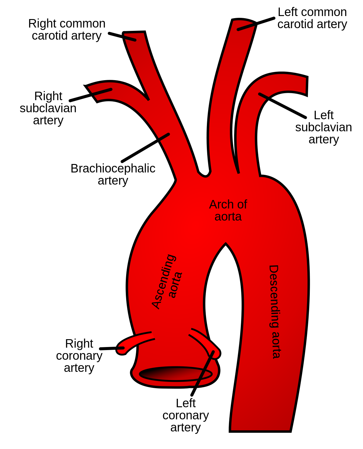
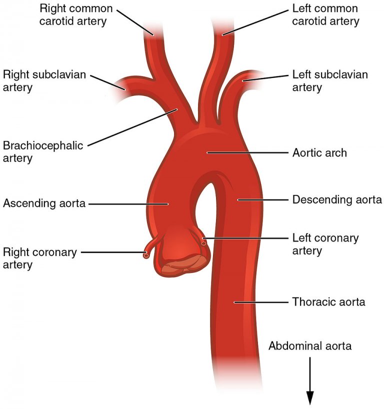
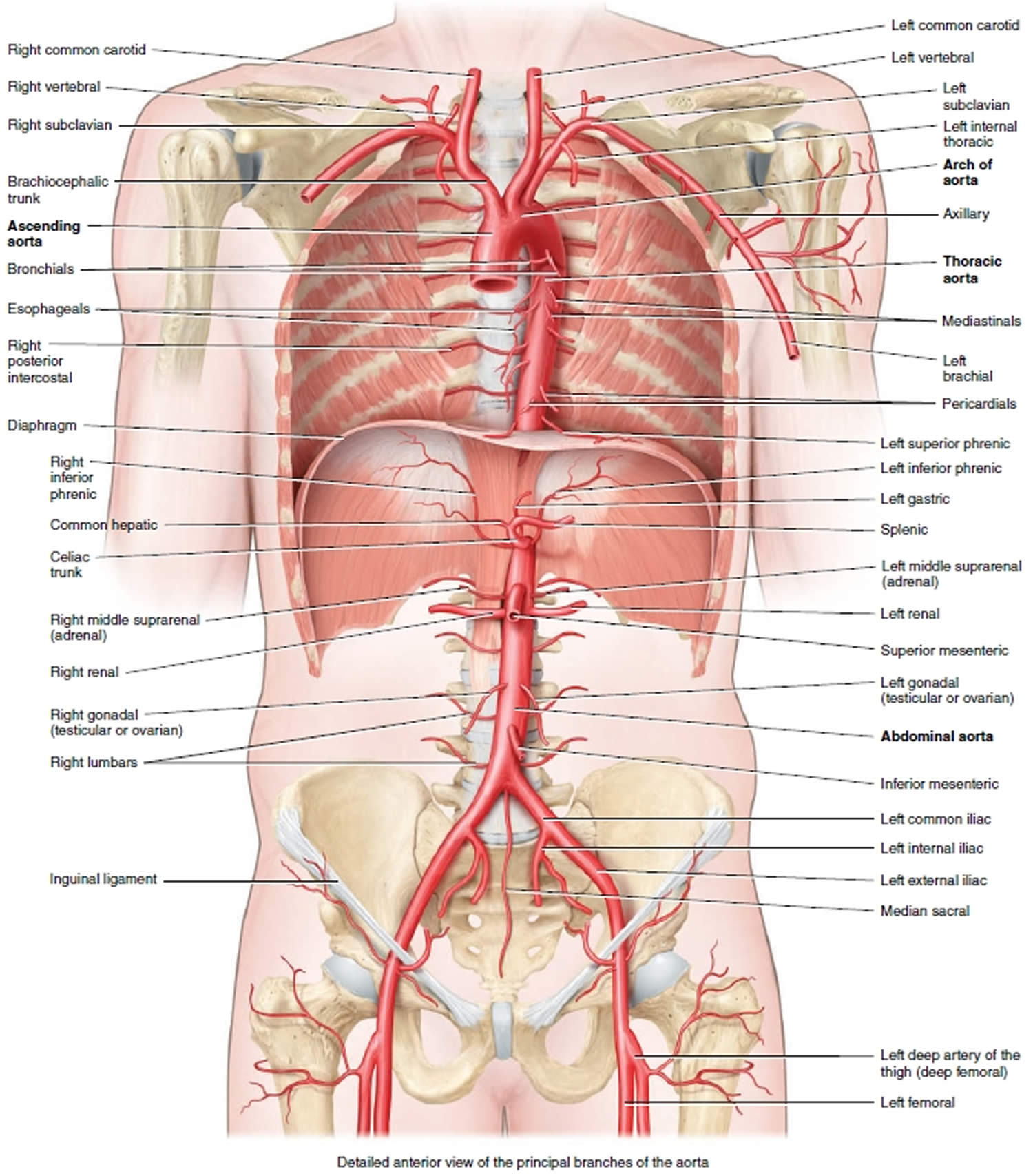
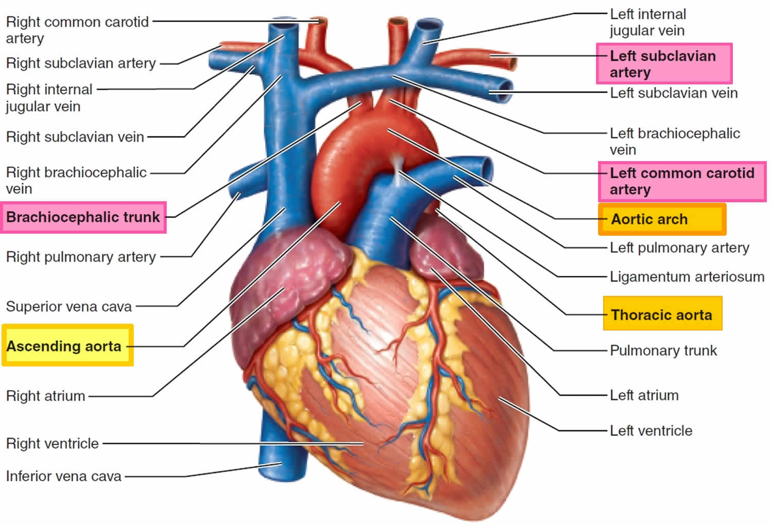
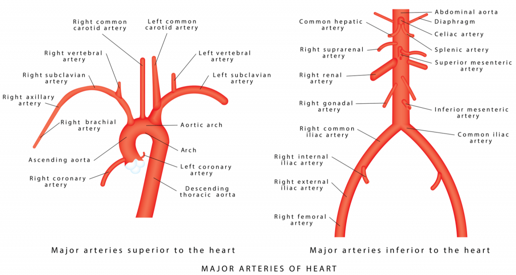
Aorta Drawing - Surgical replacement, of both the native aortic valve and ascending aorta, with an aortic homograft is an attractive therapeutic option (see below). Identify three main layers (tunics) in the wall of the aorta and their main components. The pulse of life” see an illustration of and learn about the aorta, the largest artery in the body, in the emedicinehealth image collection gallery. Web branches of the abdominal aorta / in these topics. The aorta is the largest artery in the body. Upward curve that occurs shortly after the aorta leaves the heart. Illustration of the human heart. There are two of them. The aorta supplies all of the systemic circulation, which means that the entire body, except for the respiratory zone of the lung, receives its blood from the aorta. Mild coarctation may not be diagnosed until adulthood. This sign is considered present when the posterior wall of an aortic aneurysm drapes or molds to the anterior surface of the vertebra. It is the largest artery in the body consisting of three parts that each has its special characteristics, most. Web the aortic semilunar valve is between the left ventricle and the opening of the aorta. Compare and. Illustration of the human heart. Identify three main layers (tunics) in the wall of the aorta and their main components. Web the aorta can be divided into four sections: Human heart and pulse traces. Web coarctation of the aorta symptoms depend on how much of the aorta is narrowed. It is the largest artery in the body consisting of three parts that each has its special characteristics, most. Start with the pulmonary veins. Upward curve that occurs shortly after the aorta leaves the heart. In this condition the aorta (the main artery that carries blood from the heart to the body) is narrowed or constricted. It is the largest. Rajiv verma, pediatric and adult congenital heart disease director at the children’s heart center. It is the largest artery in the body consisting of three parts that each has its special characteristics, most. The aorta is the largest artery in the body. In this condition the aorta (the main artery that carries blood from the heart to the body) is. It has three semilunar cusps/leaflets: Aorta drawing pictures, images and stock photos. Identify three main layers (tunics) in the wall of the aorta and their main components. Web branches of the abdominal aorta / in these topics. Upward curve that occurs shortly after the aorta leaves the heart. The pulse of life” see an illustration of and learn about the aorta, the largest artery in the body, in the emedicinehealth image collection gallery. This sign is considered present when the posterior wall of an aortic aneurysm drapes or molds to the anterior surface of the vertebra. Identify three main layers (tunics) in the wall of the aorta and. 18k views 2 years ago anatomy. Long, straight segment that runs from your chest (thoracic aorta) to your abdominal area (abdominal aorta). In this condition the aorta (the main artery that carries blood from the heart to the body) is narrowed or constricted. Web illustration of the abdominal aorta from the end of the thoracic aorta at the top, to. It bridges the ascending and descending aorta. Surgical replacement, of both the native aortic valve and ascending aorta, with an aortic homograft is an attractive therapeutic option (see below). Web the aorta can be divided into four sections: Relate the structural features of aorta to its function. Web the aorta, the largest artery in the body, is separated from the. Web schematic drawing of the development of the aortic arch and its branches. This sign is considered present when the posterior wall of an aortic aneurysm drapes or molds to the anterior surface of the vertebra. Illustration of the human heart. In this video, i'm taking a look at the branches of the thoracic aorta. It is the largest artery. The aorta supplies all of the systemic circulation, which means that the entire body, except for the respiratory zone of the lung, receives its blood from the aorta. It has three semilunar cusps/leaflets: Web what lily had was coarctation of the aorta, which affects one in 1,800 babies in the u.s., dr. This sign is considered present when the posterior. Most people don't have symptoms. To find a good diagram, go to google images, and type in the internal structure of the human heart. Coa can cause high blood pressure or heart damage. Identify three main layers (tunics) in the wall of the aorta and their main components. Web what lily had was coarctation of the aorta, which affects one in 1,800 babies in the u.s., dr. Mild coarctation may not be diagnosed until adulthood. Web illustration of the abdominal aorta from the end of the thoracic aorta at the top, to the bifurcation which becomes the two primitive iliac arteries. 18k views 2 years ago anatomy. Web the aorta is divided into three parts: Web coarctation of the aorta symptoms depend on how much of the aorta is narrowed. Surgical replacement, of both the native aortic valve and ascending aorta, with an aortic homograft is an attractive therapeutic option (see below). Illustration of the human heart. Learn the structure, location in the human heart, anatomy, various functions with labelled diagrams. Web what is aorta? It terminates at the level of l4 by bifurcating into the left and right common iliac arteries. Find a piece of paper and something to draw with.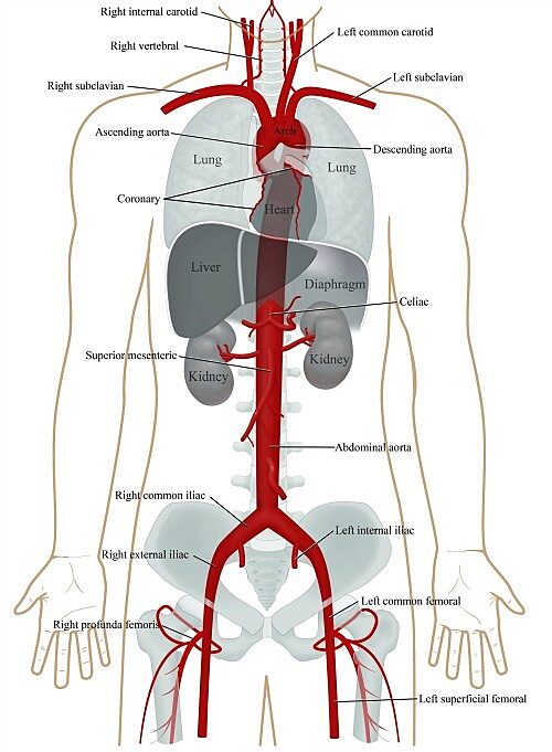
Aorta anatomy UF Health
Ascending aorta Wikipedia
Aorta Explained Anatomy 101 For patients
Aorta anatomy, function, branches, location & aorta problems
Aorta anatomy, function, branches, location & aorta problems
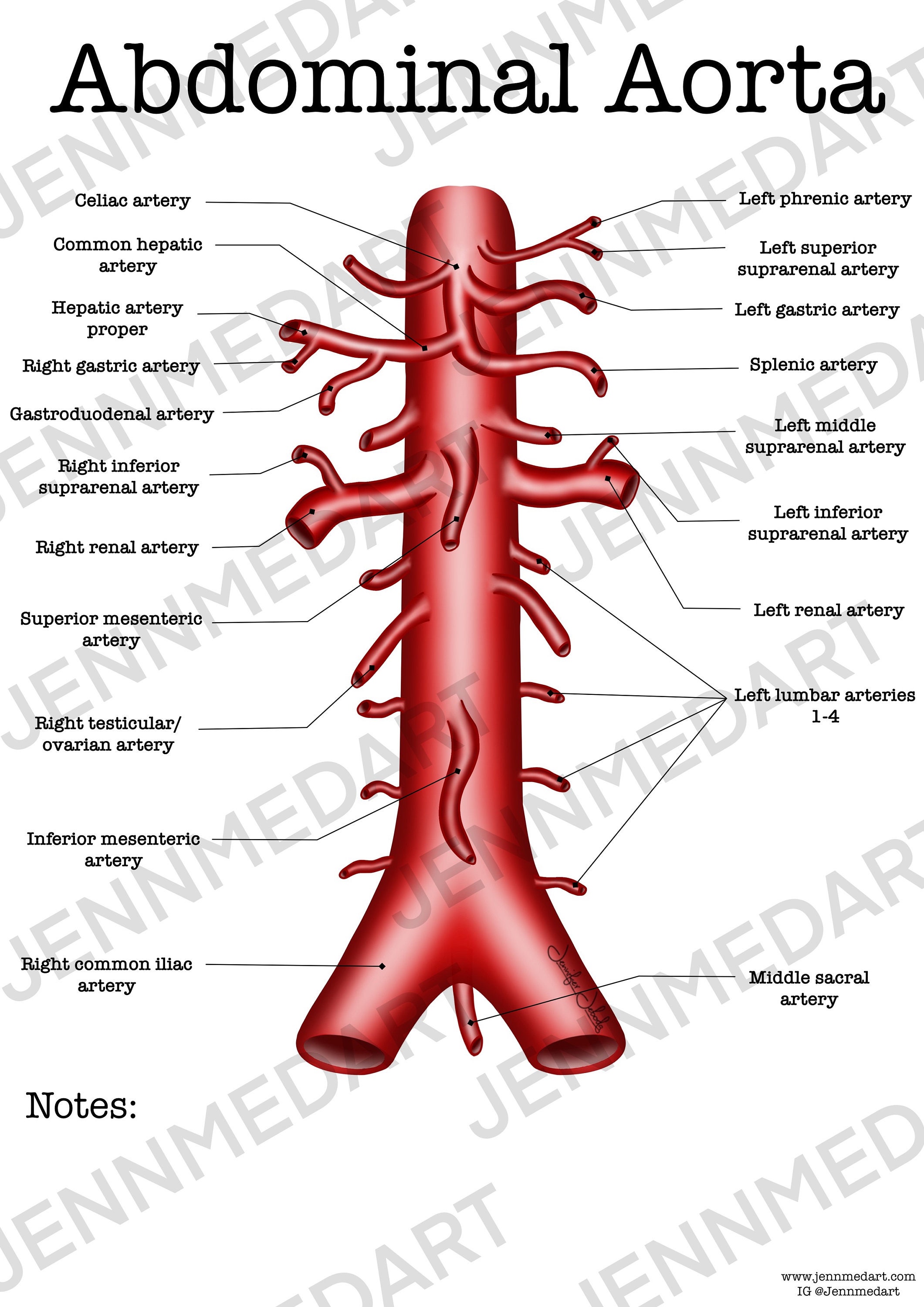
Abdominal Aorta Anatomy Worksheet Single FILLED Digital Download
.png)
Radiopaedia Drawing Aortic arch and its branches English labels

Aorta Drawing Stock Image C020/9510 Science Photo Library

Aorta wikidoc
Aorta Explained Anatomy 101 For patients
Human Heart And Pulse Traces.
They Will Be To The Lower Left Of The Aorta.
The Ascending Aorta (Where The Aorta Initially Leaves The Heart And Points Toward The Head), The Arch Of The Aorta (Where The Aorta Changes Direction), And The Descending Aorta (Where The Aorta Points Toward The Feet).
Compare And Contrast The Main Histological Features Of Tissues That Can Be Identified Via H&E And Elastic/Trichrome Stains.
Related Post: